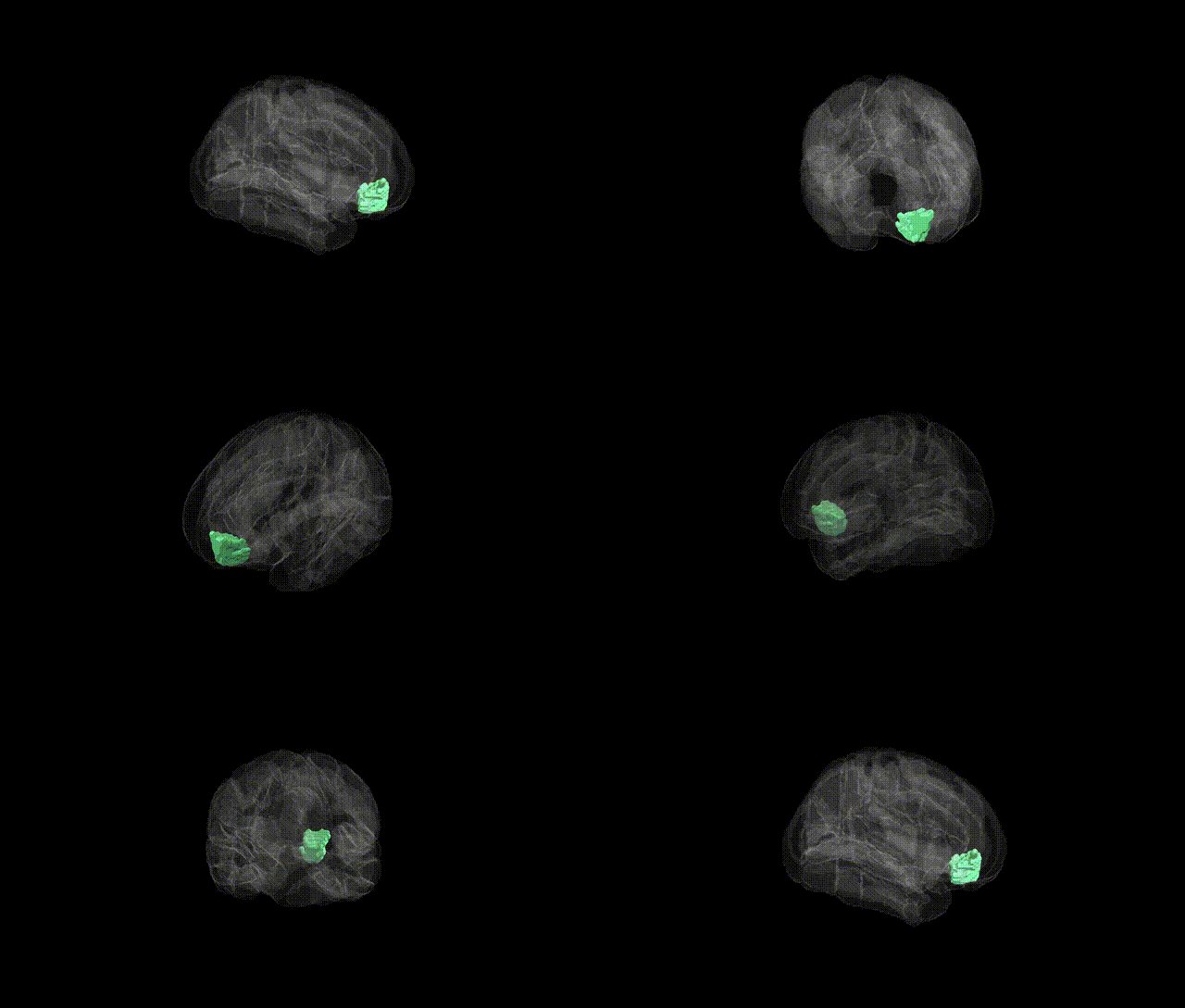
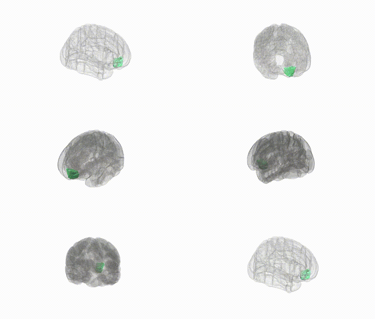
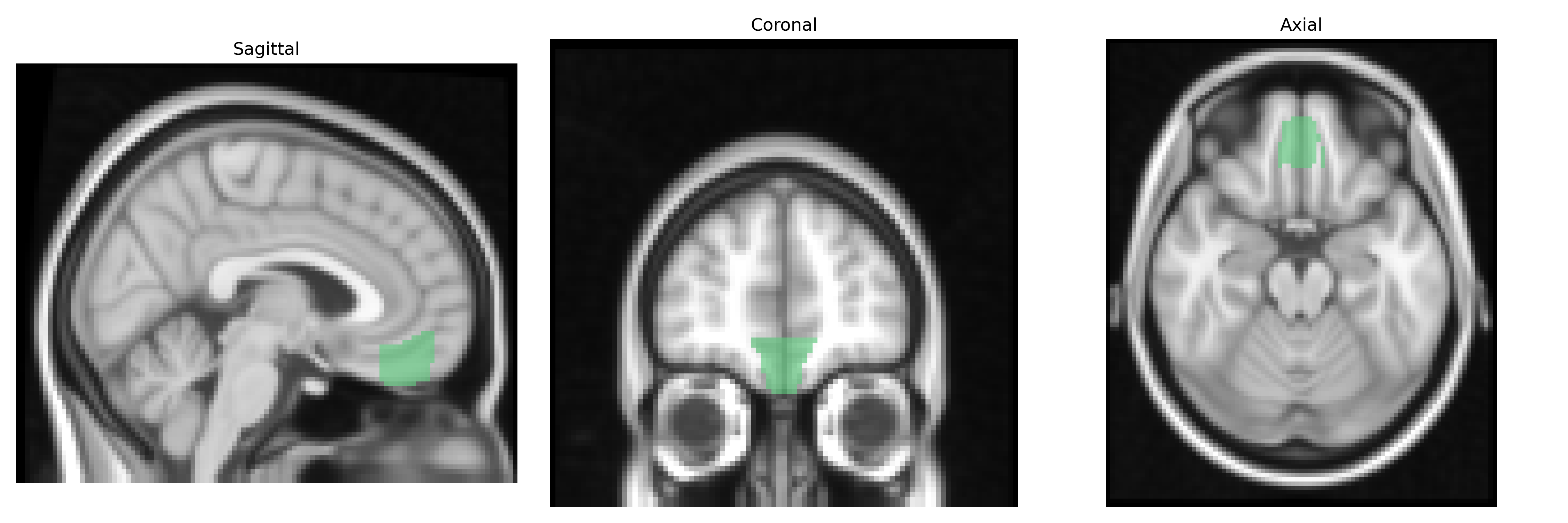
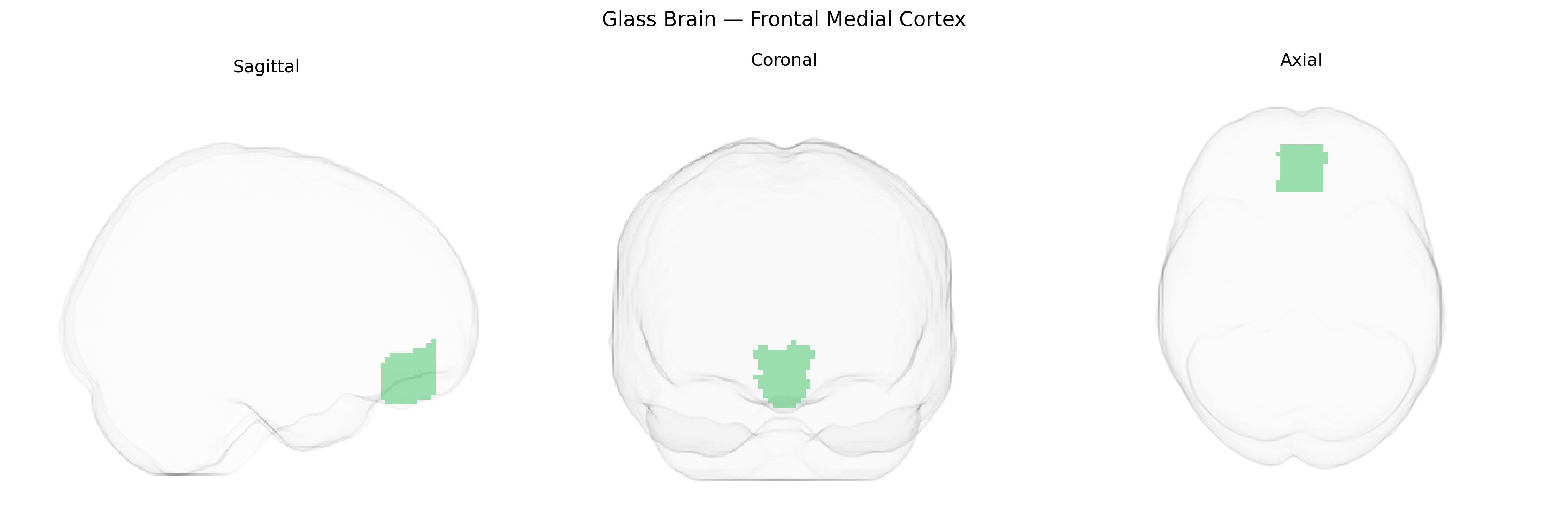
The bilateral frontal medial cortex, as defined in the Harvard-Oxford cortical atlas (maxprob, thr25, 2 mm), encompasses medial portions of the frontal lobe along the interhemispheric fissure, including regions commonly associated with the medial prefrontal cortex. This area participates in higher-order executive functions, valuation and decision-making processes, social and self-referential cognition, and regulation of affect and motivation, integrating information from limbic, parietal, and subcortical structures. It forms part of large-scale networks such as the default mode network and fronto-limbic circuits, and is implicated in neuropsychiatric conditions including depression, anxiety disorders, and schizophrenia. There is no direct Wikipedia article specifically titled “Frontal Medial Cortex”; a closely related and encompassing structure is the Medial prefrontal cortex.
The bilateral frontal medial cortex (often approximating medial prefrontal and anterior cingulate regions in the Harvard–Oxford Cortical atlas) has been repeatedly implicated in genetic studies of brain structure, function, and neuropsychiatric risk. GWAS of cortical thickness, surface area, and volume have identified associations between frontal medial measures and variants in genes involved in neurodevelopment, synaptic function, and cell adhesion (for example, loci near genes such as PAX6, TBR1, and various cadherins and protocadherins), with some findings overlapping polygenic architectures for general cognitive ability and educational attainment. Imaging genetics studies link polygenic risk scores for major depressive disorder, schizophrenia, bipolar disorder, autism spectrum disorder, and ADHD with altered medial frontal morphology and activity, particularly in tasks involving reward processing, self-referential thought, and cognitive control. Risk alleles in regulatory regions affecting dopaminergic and serotonergic signaling (e.g., near DRD2, SLC6A4) and glutamatergic genes (e.g., GRM3, GRIN2A) have been associated with functional differences in medial frontal networks, while APOE and other Alzheimer’s disease–related variants show relationships with medial frontal atrophy and connectivity in aging and preclinical dementia. Large consortia (ENIGMA, UK Biobank, and others) collectively support that genetic variation contributes substantially to individual differences in frontal medial structure and function, with this region serving as a key intermediate phenotype linking common variants to mood, psychotic, anxiety, and substance use disorders, as well as personality traits such as neuroticism and impulsivity.
Overview generated by GPT-4o (2026).
Region ID: 25
Hemisphere: bilateral
Atlas: HarvardOxford cort maxprob thr25 2mm
Full Quality Version: Download MP4
Full Quality Version: Download MP4
Wali Sidiqyar*, Gaurav Rudravaram*, Elyssa M. McMaster, Trent M. Schwartz, Adam M. Saunders, Kurt G. Schilling, Bennett A. Landman "Introducing SPINS: A Shared Public Visualization Library of Neuroanatomical Structures." Medical Imaging with Deep Learning- short paper
This resource is licensed under CC0 1.0 Universal (Public Domain).