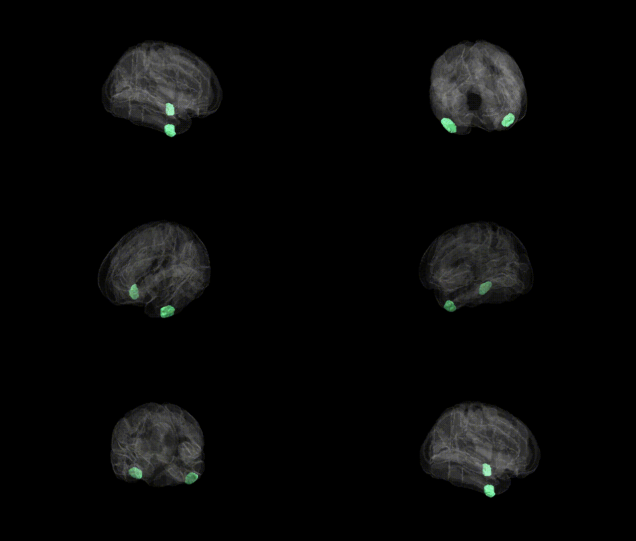
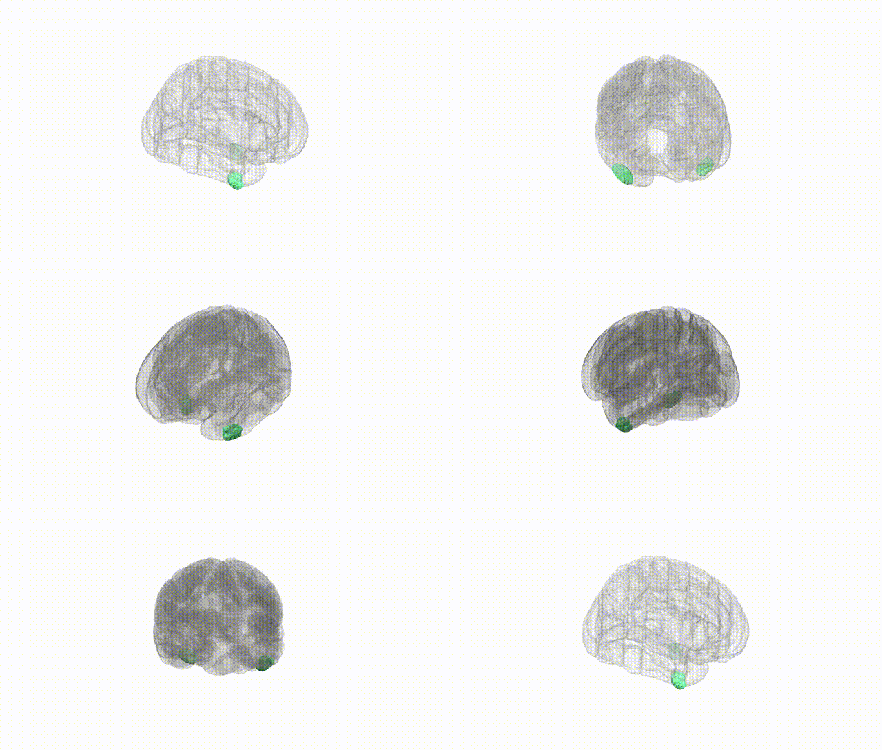
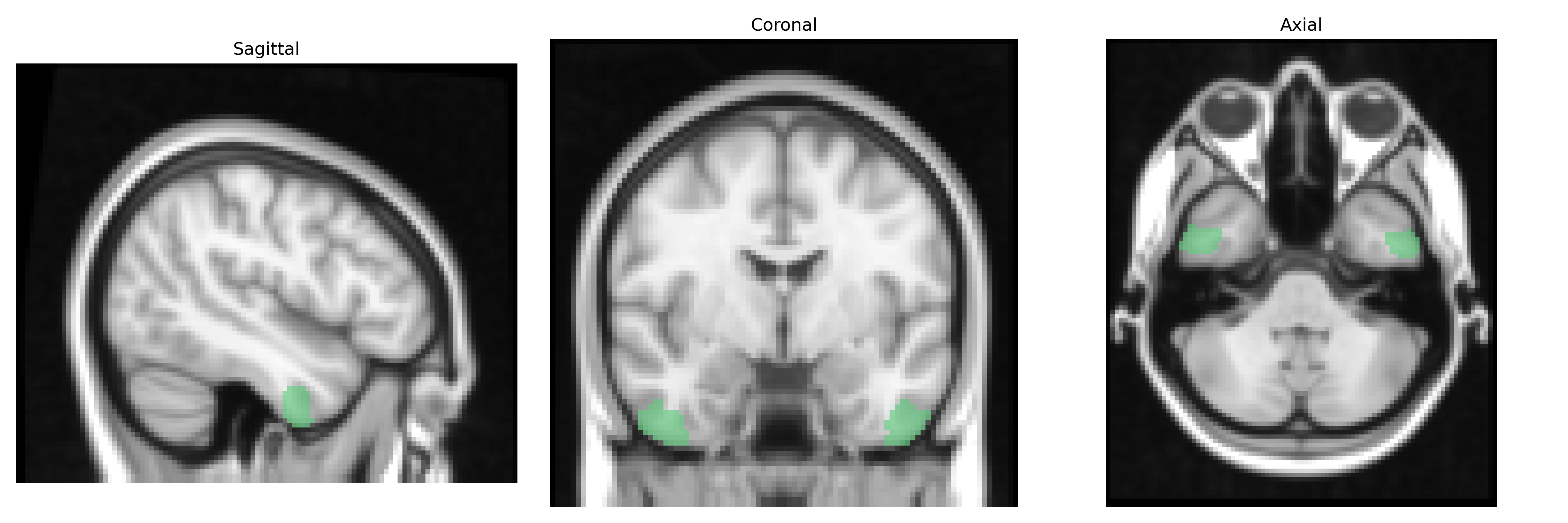
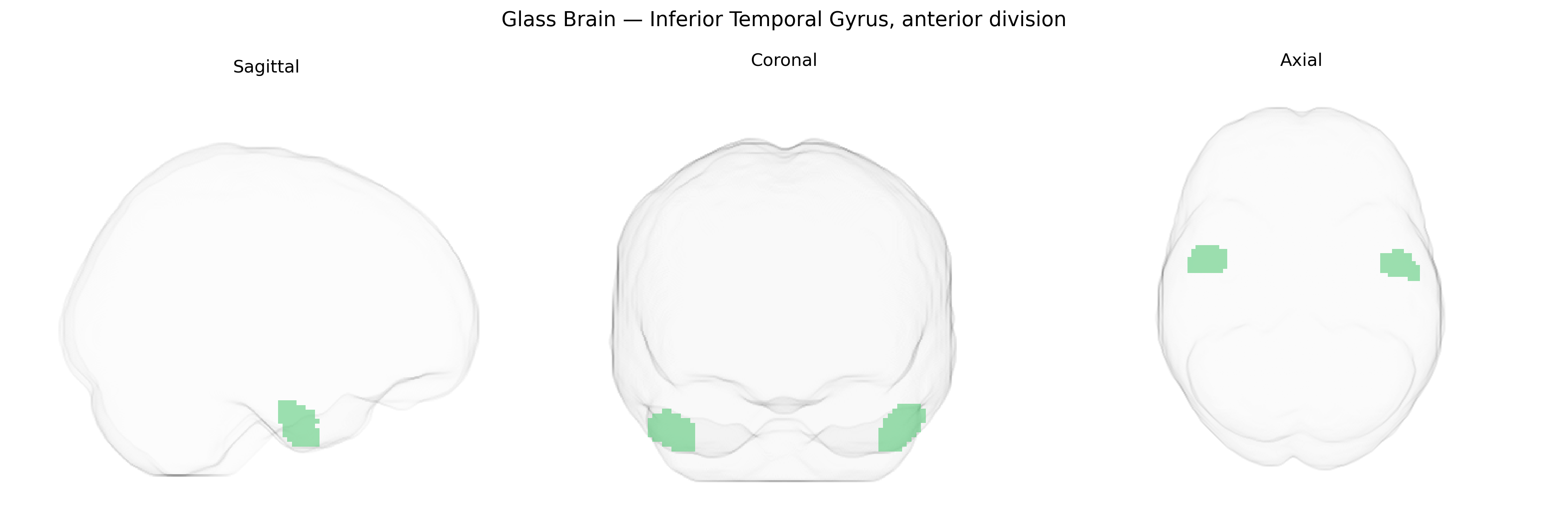
The bilateral Inferior Temporal Gyrus, anterior division (Harvard-Oxford Cortical Atlas, maxprob thr25, 2 mm) corresponds to the rostral portion of the inferior temporal cortex along the ventral lateral surface of the temporal lobe, extending anteriorly from the mid-portion of the gyrus toward the temporal pole. This region lies inferior to the middle temporal gyrus and lateral to the fusiform gyrus, with cytoarchitectonic and functional properties associated with high-level visual processing, including object and form recognition, complex feature integration, and participation in the ventral “what” visual pathway. It receives convergent input from earlier visual areas and contributes to the transformation of visual information into semantic and categorical representations. In humans, this territory has been implicated by neuroimaging and lesion studies in the recognition of objects and faces, as well as in certain aspects of visual memory and semantic processing. There is no direct link for the “Inferior Temporal Gyrus, anterior division” subdivision; a closely related structure is the Inferior temporal gyrus.
The bilateral anterior division of the Inferior Temporal Gyrus (ITG) has been implicated in genetic studies primarily through imaging genetics and GWAS of cortical structure rather than direct locus-specific association. Large-scale neuroimaging GWAS, such as those from ENIGMA and UK Biobank, have identified multiple common variants influencing cortical thickness and surface area in temporal regions that encompass the ITG, with notable contributions from loci near genes involved in neurodevelopment, synaptic function, and axon guidance (e.g., variants near genes such as DAAM1, PPP1R17, and others reported in global temporal cortex measures). ITG structure and function are genetically correlated with higher-order visual processing, semantic cognition, and face/object recognition, and genetic influences on these phenotypes (including variants in KIAA0319, CNTNAP2, and FOXP2 for language/reading; and genes implicated in prosopagnosia and autism spectrum traits) have been linked to altered activation or morphology in inferior temporal regions, though not always specifically parsed to the anterior division. GWAS of psychiatric and neurodevelopmental disorders—such as schizophrenia, autism spectrum disorder, and major depression—show polygenic risk scores that relate to structural and functional changes in inferior temporal and adjacent ventral temporal cortex, suggesting that widely distributed risk variants (e.g., in synaptic genes like CACNA1C, GRIN2A, and others) contribute to ITG alterations as part of broader network-level effects. However, current evidence supports a polygenic, distributed architecture, and no single gene or variant is uniquely or specifically established as acting on the bilateral anterior ITG in isolation; rather, genetic influences on this region reflect shared neurodevelopmental and cognitive pathways affecting temporal lobe organization more generally.
Overview generated by GPT-4o (2026).
Region ID: 14
Hemisphere: bilateral
Atlas: HarvardOxford cort maxprob thr25 2mm
Full Quality Version: Download MP4
Full Quality Version: Download MP4
Wali Sidiqyar*, Gaurav Rudravaram*, Elyssa M. McMaster, Trent M. Schwartz, Adam M. Saunders, Kurt G. Schilling, Bennett A. Landman "Introducing SPINS: A Shared Public Visualization Library of Neuroanatomical Structures." Medical Imaging with Deep Learning- short paper
This resource is licensed under CC0 1.0 Universal (Public Domain).