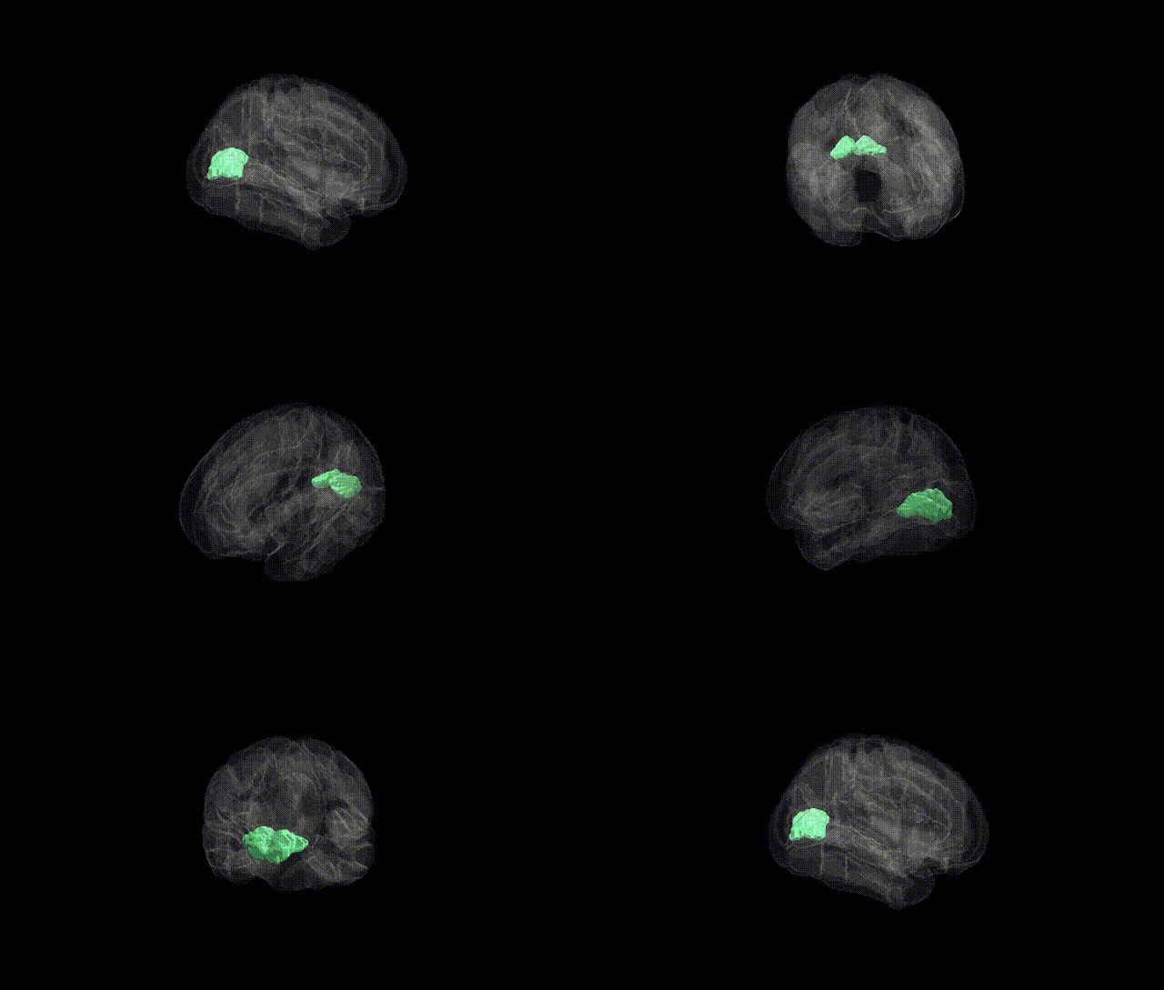
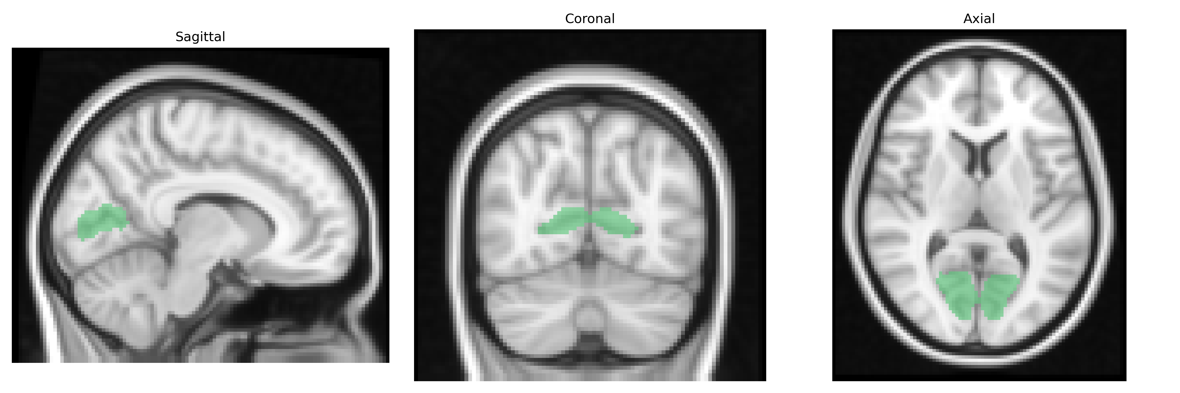
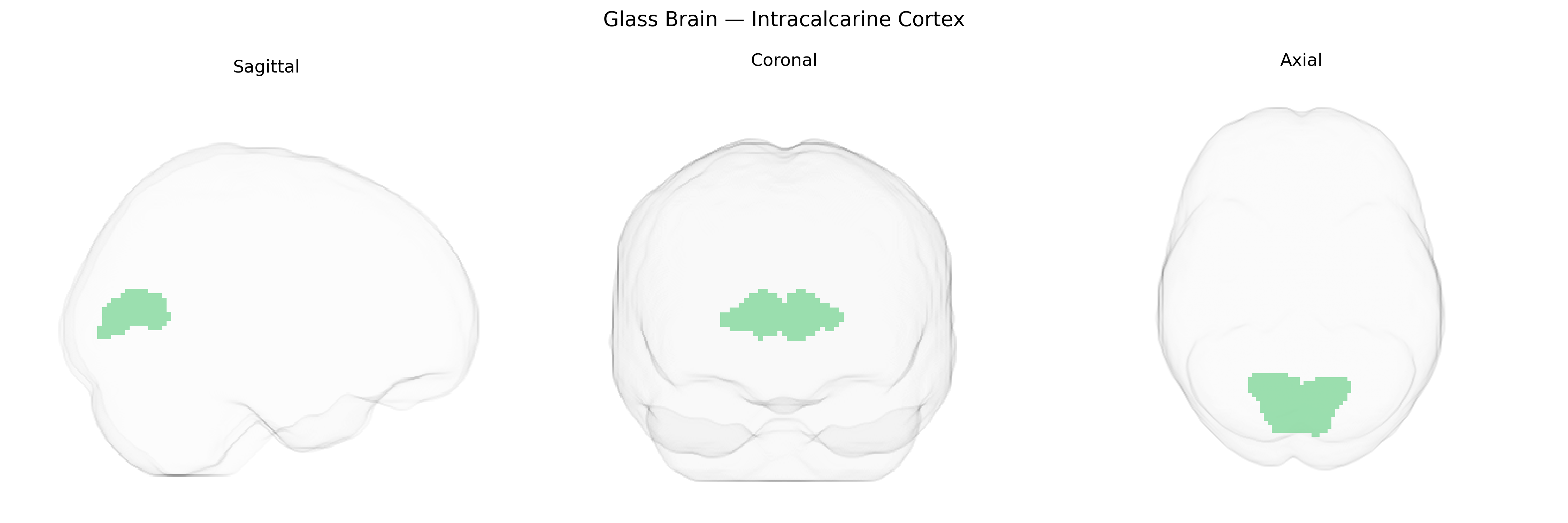
The bilateral Intracalcarine Cortex corresponds primarily to the primary visual cortex (V1) located along the calcarine sulcus in the occipital lobe, as defined in the Harvard-Oxford cortical structural atlas. This region is the principal recipient of visual input from the lateral geniculate nucleus of the thalamus and is organized retinotopically, with distinct portions representing specific regions of the visual field. Neurons in this cortex are highly sensitive to basic visual features such as orientation, spatial frequency, contrast, and ocular dominance, forming the foundation for subsequent visual processing in higher-order visual areas. Functionally, the Intracalcarine Cortex is essential for conscious visual perception and damage to this region can lead to characteristic visual field deficits (scotomas) or cortical blindness. There is no direct Wikipedia article for Intracalcarine Cortex; a closely related and encompassing structure is Primary visual cortex.
The bilateral intracalcarine cortex, corresponding largely to primary visual cortex (V1) in the Harvard–Oxford Cortical Atlas, has been implicated in several genetic and GWAS-based findings, primarily through imaging genetics studies that link common variants to cortical thickness, surface area, and functional activation in visual regions. Large-scale neuroimaging GWAS (e.g., ENIGMA, UK Biobank) have identified multiple loci associated with occipital and calcarine cortical measures, including variants near genes involved in neurodevelopment, synaptic function, and axon guidance (such as those in Wnt signaling and cell adhesion pathways), although most associations are regionally broad and not specific to intracalcarine cortex alone. Polygenic risk scores for schizophrenia, major depressive disorder, bipolar disorder, and autism spectrum conditions have shown correlations with structural and functional alterations in occipital and calcarine regions, suggesting shared genetic architectures that influence early visual processing and higher-order perceptual functions. In addition, GWAS of visual acuity and refractive error (myopia, hyperopia) have reported associations with genes that modulate visual pathway development and cortical plasticity, indirectly implicating calcarine visual cortex structure and function. However, as of current literature, there are no strongly replicated, highly specific genetic variants uniquely tied to the intracalcarine cortex independent of broader occipital or global brain measures, and most genetic links to this region arise from distributed neurodevelopmental and psychiatric risk architectures that affect multiple cortical networks including primary visual areas.
Overview generated by GPT-4o (2026).
Region ID: 24
Hemisphere: bilateral
Atlas: HarvardOxford cort maxprob thr25 2mm
Full Quality Version: Download MP4
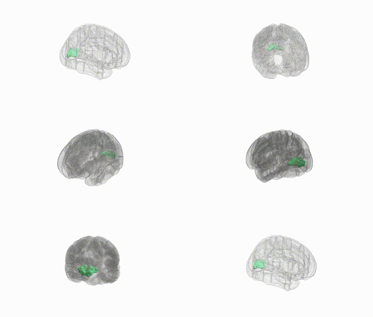
Full Quality Version: Download MP4
Wali Sidiqyar*, Gaurav Rudravaram*, Elyssa M. McMaster, Trent M. Schwartz, Adam M. Saunders, Kurt G. Schilling, Bennett A. Landman "Introducing SPINS: A Shared Public Visualization Library of Neuroanatomical Structures." Medical Imaging with Deep Learning- short paper
This resource is licensed under CC0 1.0 Universal (Public Domain).