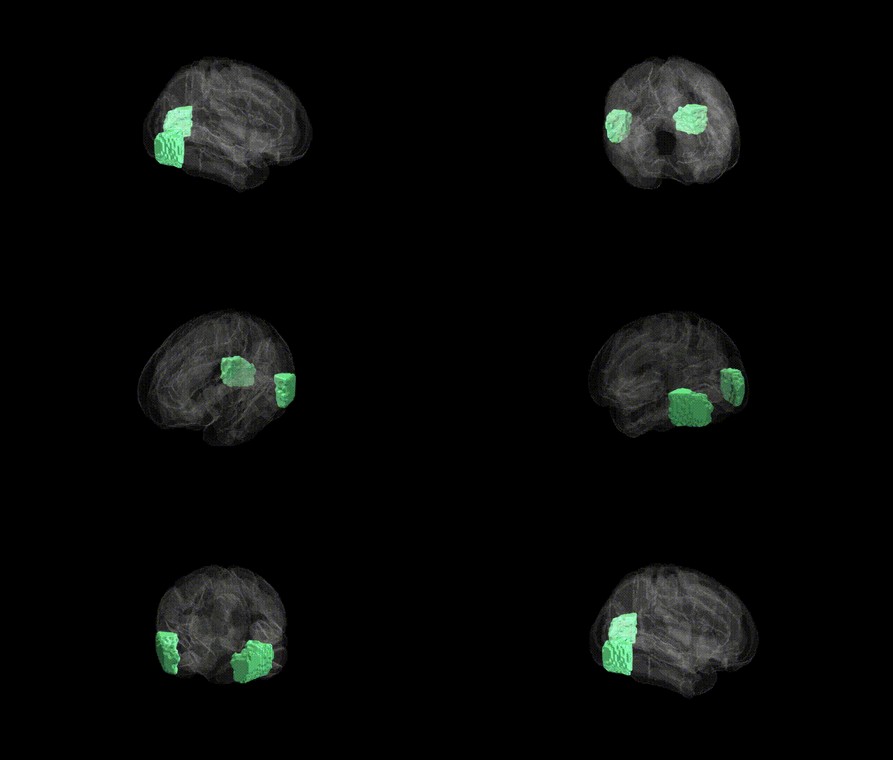
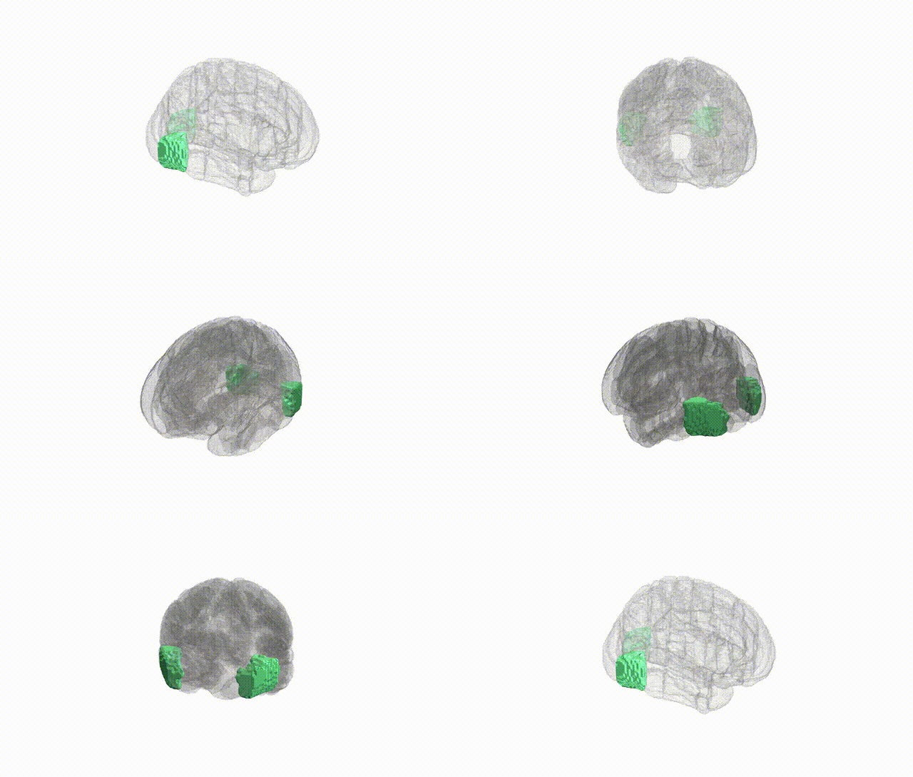
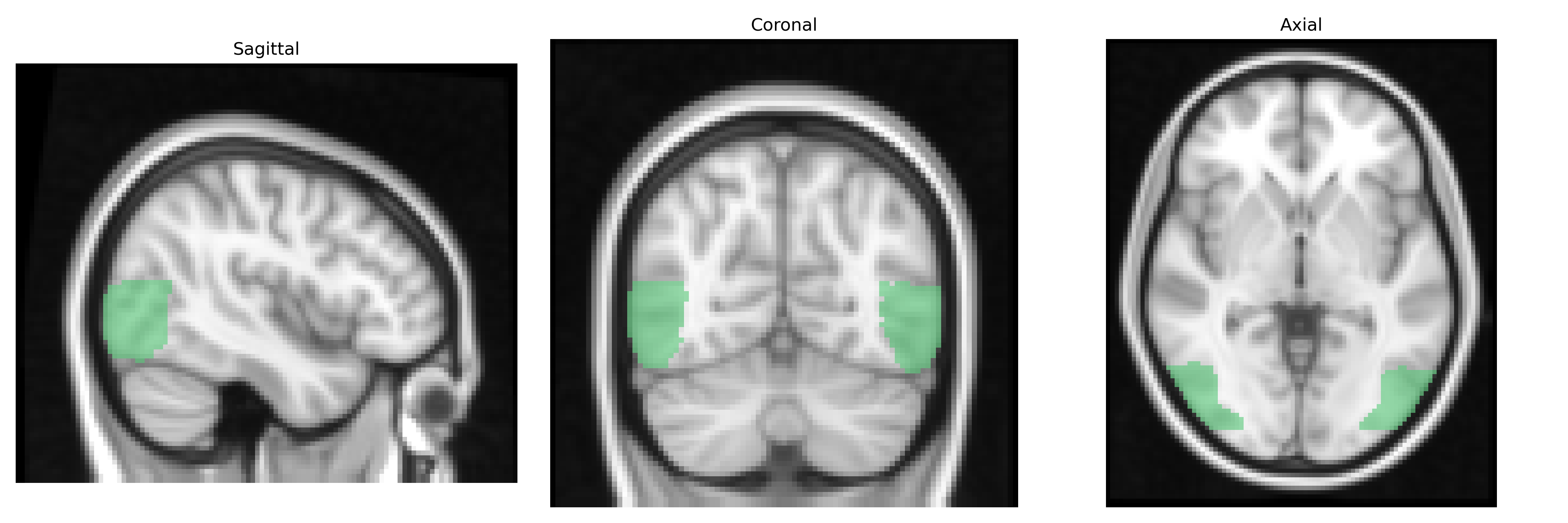
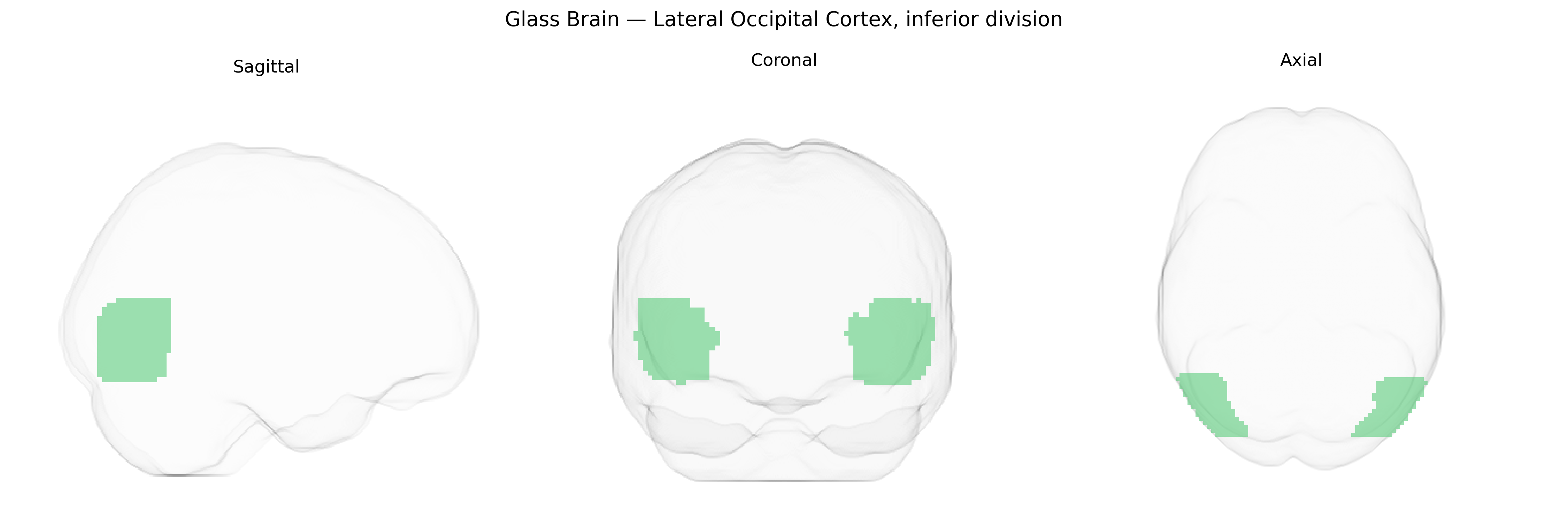
The bilateral Lateral Occipital Cortex, inferior division, as defined in the Harvard-Oxford cortical maxprob atlas, comprises ventrolateral portions of the occipital lobe situated adjacent to the temporal–occipital border and inferior to the superior division of the lateral occipital cortex. This region participates in higher-order visual processing, particularly object shape and form recognition, integrating information from primary and secondary visual areas and contributing to the ventral “what” visual pathway. It is implicated in visual object perception, including recognition of complex shapes and possibly faces and tools, with bilateral organization supporting robust and redundant visual representations across hemispheres. There is no direct Wikipedia article for this specific inferior division; a related entry is Lateral occipital cortex.
The bilateral lateral occipital cortex, inferior division—corresponding to ventral occipital/occipitotemporal visual areas in the Harvard–Oxford atlas—has been implicated in several genetic and GWAS-based findings, largely through imaging genetics and large-scale consortia such as ENIGMA and UK Biobank. Structural and functional measures in this region (e.g., cortical thickness, surface area, activation during visual and face processing tasks) show significant heritability and have been associated with common genetic variants near or within genes involved in neurodevelopment, synaptic function, and axon guidance, including loci around genes such as MEF2C, DCC, and variants in pathways regulating visual cortical development and plasticity. GWAS of occipital cortical morphology have identified risk alleles overlapping with those for cognitive ability and educational attainment, suggesting shared genetic architecture between visual association cortex structure and higher-order cognition. In disorders, imaging genetics studies of schizophrenia, autism spectrum disorder, dyslexia, and major depressive disorder have reported genotype-dependent differences in lateral occipital cortex structure or activation—for example, polygenic risk scores for schizophrenia and autism correlating with altered occipital cortical thickness or connectivity, and dyslexia-associated variants in genes such as KIAA0319 and DCDC2 relating to atypical occipitotemporal activation during reading. Additionally, GWAS of face recognition ability and visual perceptual traits have implicated this region’s activation patterns and structure, consistent with its role in object and face processing and the broader genetic architecture of visual cognition.
Overview generated by GPT-4o (2026).
Region ID: 23
Hemisphere: bilateral
Atlas: HarvardOxford cort maxprob thr25 2mm
Full Quality Version: Download MP4
Full Quality Version: Download MP4
Wali Sidiqyar*, Gaurav Rudravaram*, Elyssa M. McMaster, Trent M. Schwartz, Adam M. Saunders, Kurt G. Schilling, Bennett A. Landman "Introducing SPINS: A Shared Public Visualization Library of Neuroanatomical Structures." Medical Imaging with Deep Learning- short paper
This resource is licensed under CC0 1.0 Universal (Public Domain).