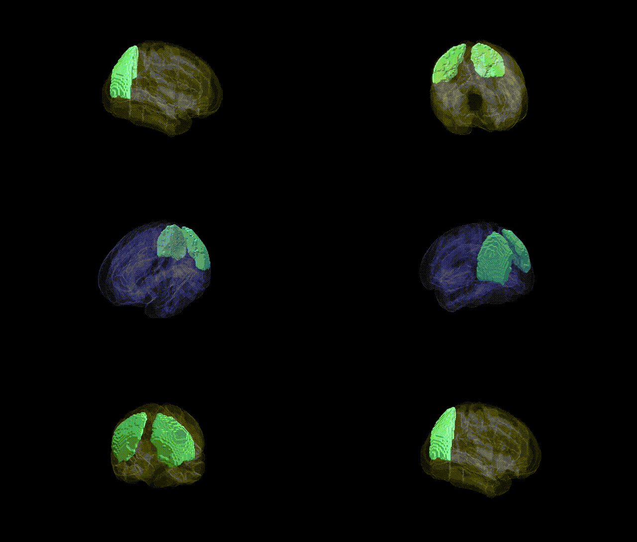
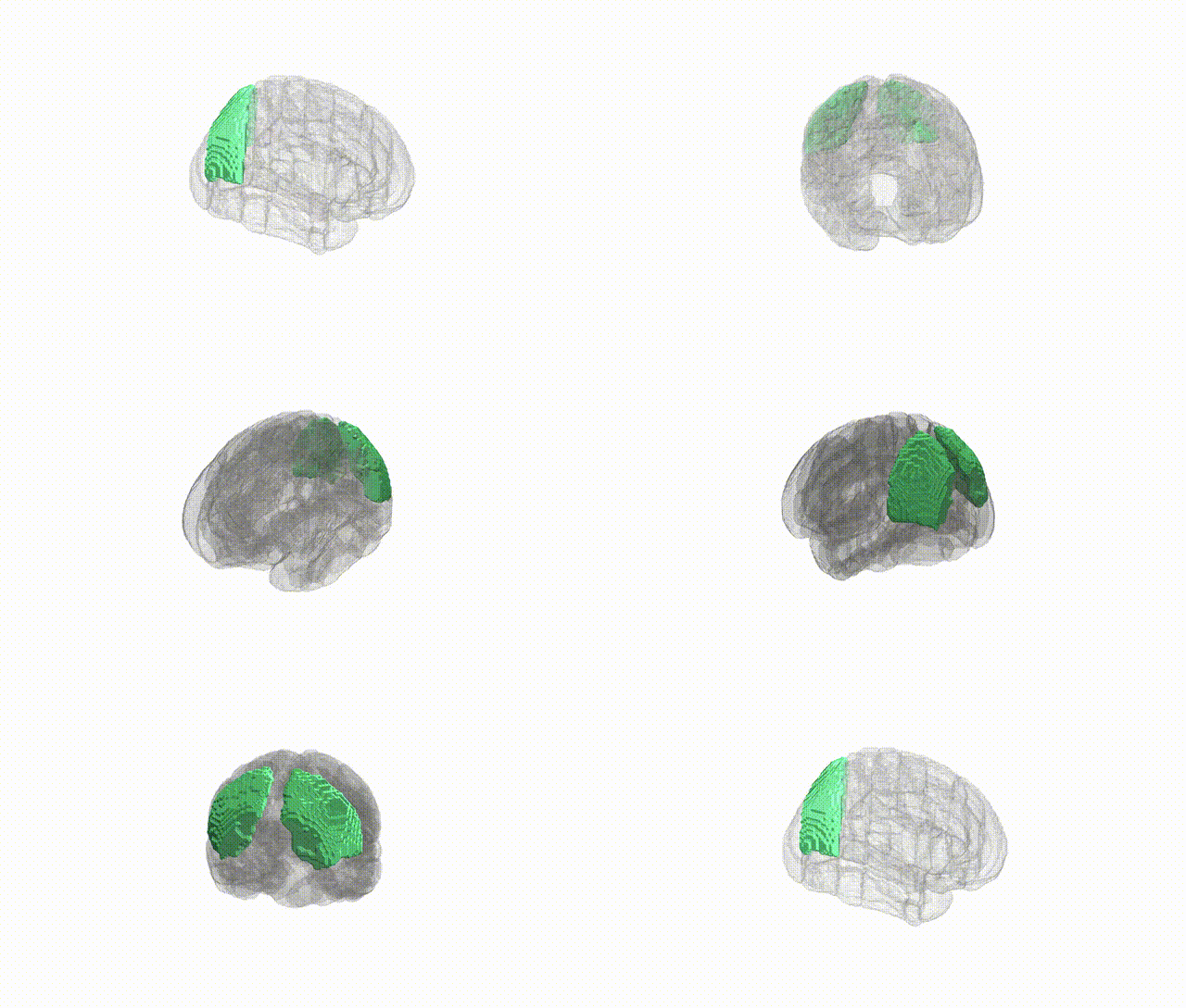
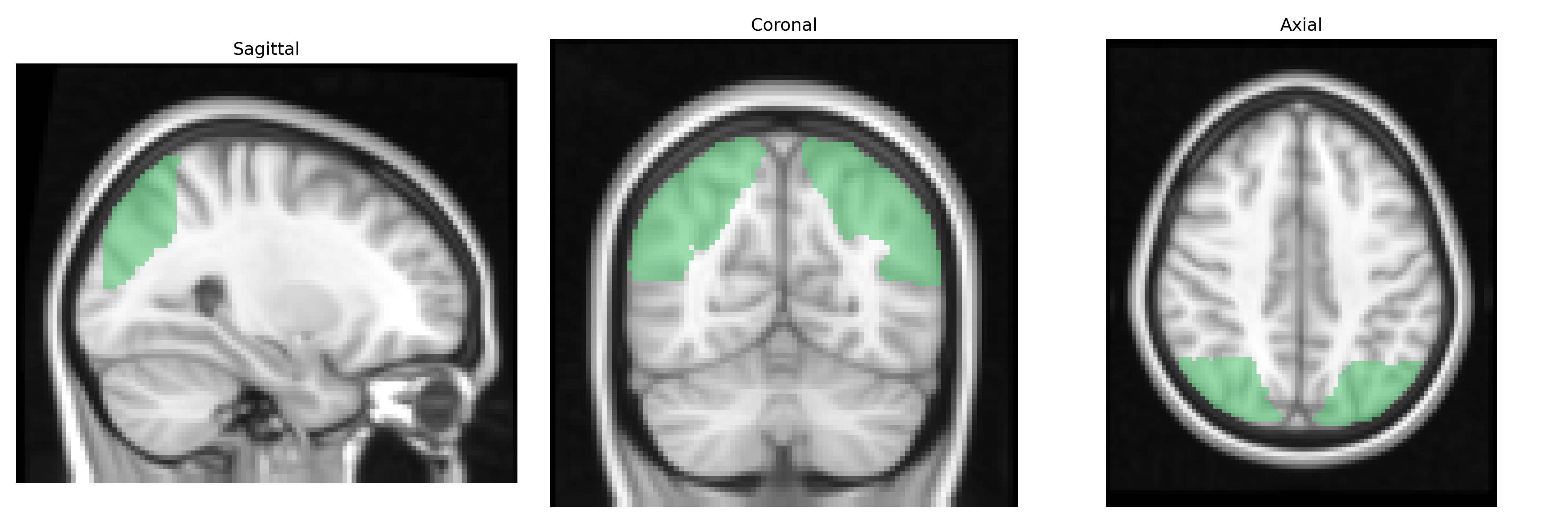
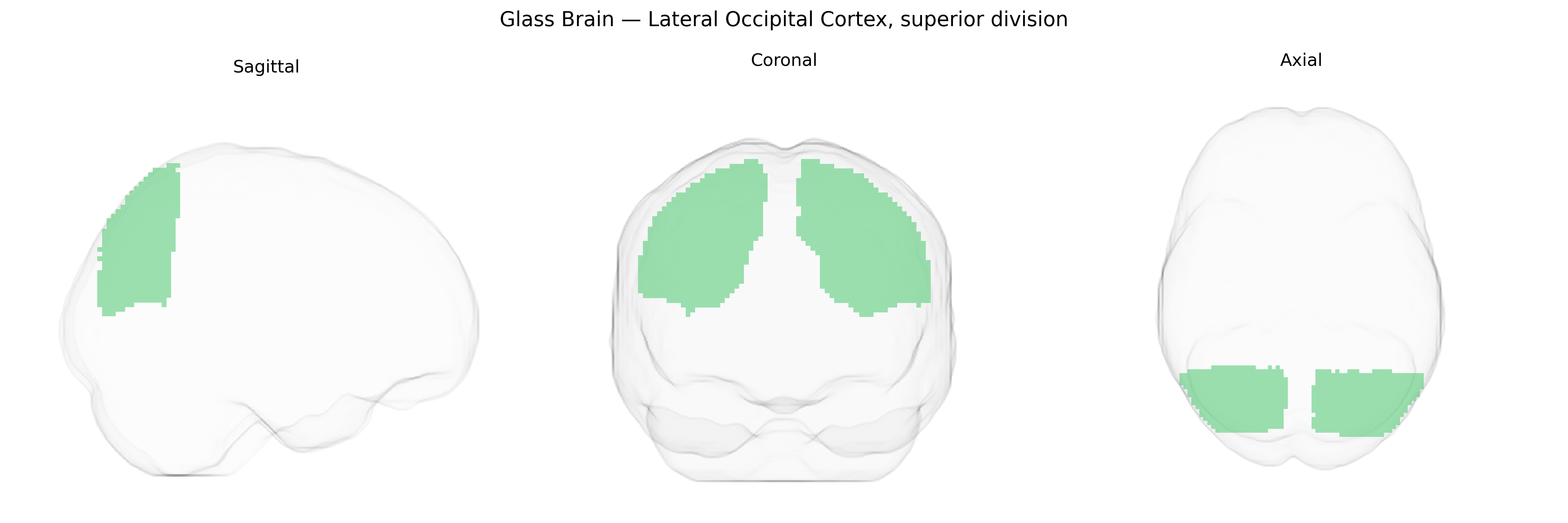
The bilateral Lateral Occipital Cortex, superior division, as defined in the Harvard-Oxford cortical maximum probability atlas (25% threshold, 2 mm), corresponds to a dorsal portion of the lateral occipital lobe situated posterior to the parietal cortex and superior to the inferior lateral occipital regions. This region participates prominently in higher-order visual processing, including shape, object, and complex form perception, and is often considered part of the lateral occipital complex involved in object recognition. It receives convergent input from earlier visual areas and contributes to the integration of visual features into coherent percepts, interfacing with dorsal and ventral visual streams. There is no direct Wikipedia article for this exact atlas-defined subdivision; a closely related structure is the Lateral occipital cortex.
The bilateral lateral occipital cortex, superior division (as defined in the Harvard–Oxford cortical atlas) has been implicated in several imaging genetics and GWAS findings related to visual processing, cortical morphology, and neuropsychiatric liability. Large-scale brain-structure GWAS (e.g., ENIGMA and UK Biobank–based studies) have identified associations between common variants in genes involved in neurodevelopment and synaptic function—such as those in or near MAPT, HMGA2, and various glutamatergic and axon-guidance loci—and occipital cortical thickness or surface area, including regions overlapping the lateral occipital cortex. Polygenic risk scores for schizophrenia, bipolar disorder, and major depression show correlations with structural alterations in lateral occipital areas, while autism spectrum disorder and dyslexia genetics have been linked to atypical occipital activation during visual and language tasks. Variants in genes affecting visual system development (for example, in pathways related to axon guidance, myelination, and visual phototransduction) have also been associated with occipital lobe volume or function in GWAS of visual acuity and visual cortex metrics. Although few studies target the “lateral occipital cortex, superior division” label specifically, convergent genetic evidence points to this region as a downstream locus where polygenic influences on cortical development, visual perception, and psychiatric risk manifest in measurable structural and functional differences.
Overview generated by GPT-4o (2026).
Region ID: 22
Hemisphere: bilateral
Atlas: HarvardOxford cort maxprob thr25 2mm
Full Quality Version: Download MP4
Full Quality Version: Download MP4
Wali Sidiqyar*, Gaurav Rudravaram*, Elyssa M. McMaster, Trent M. Schwartz, Adam M. Saunders, Kurt G. Schilling, Bennett A. Landman "Introducing SPINS: A Shared Public Visualization Library of Neuroanatomical Structures." Medical Imaging with Deep Learning- short paper
This resource is licensed under CC0 1.0 Universal (Public Domain).