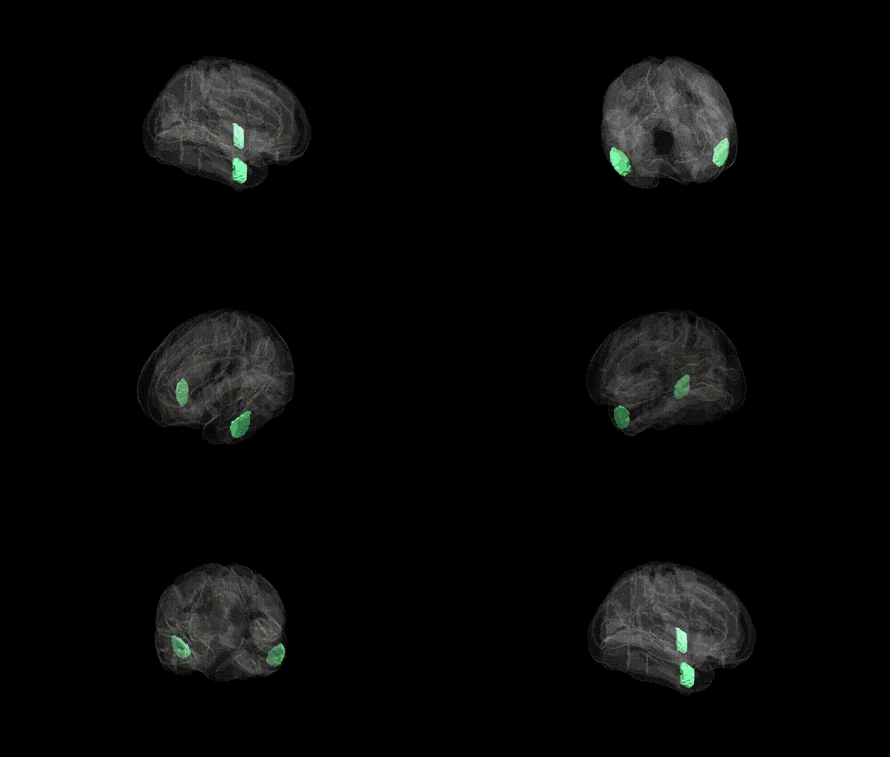
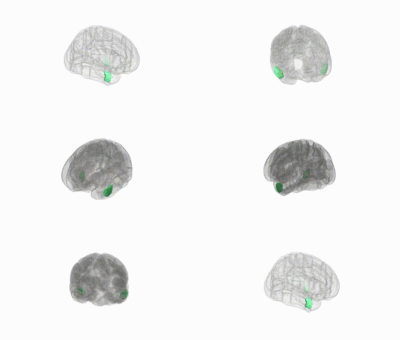
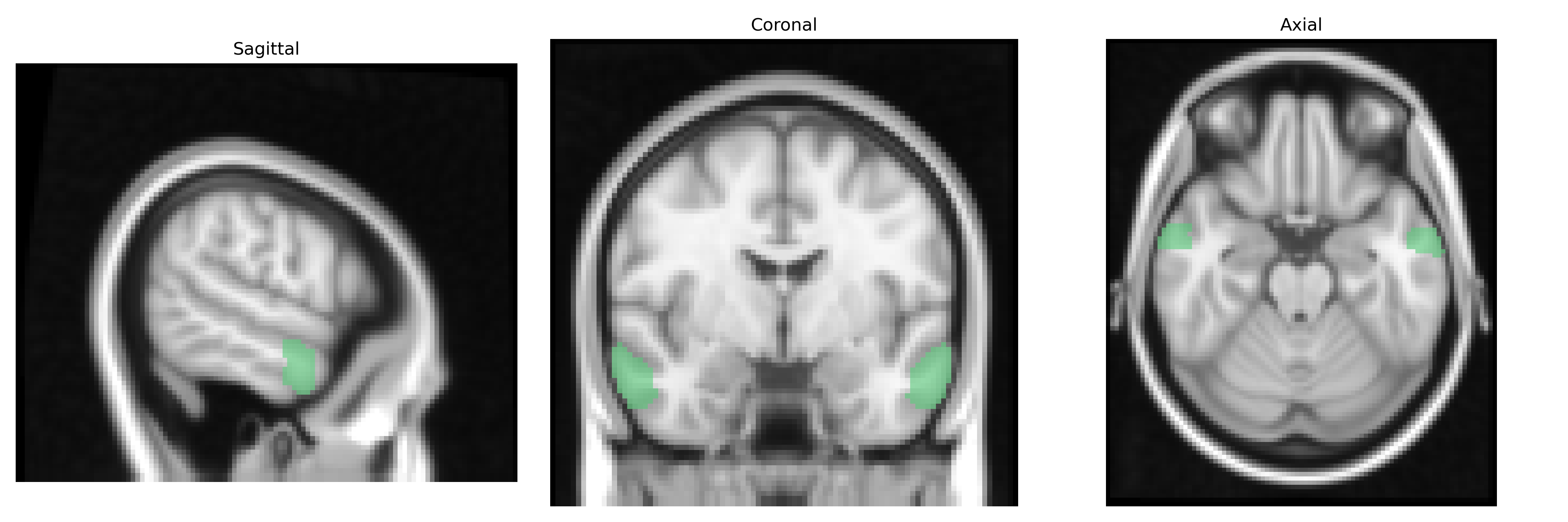
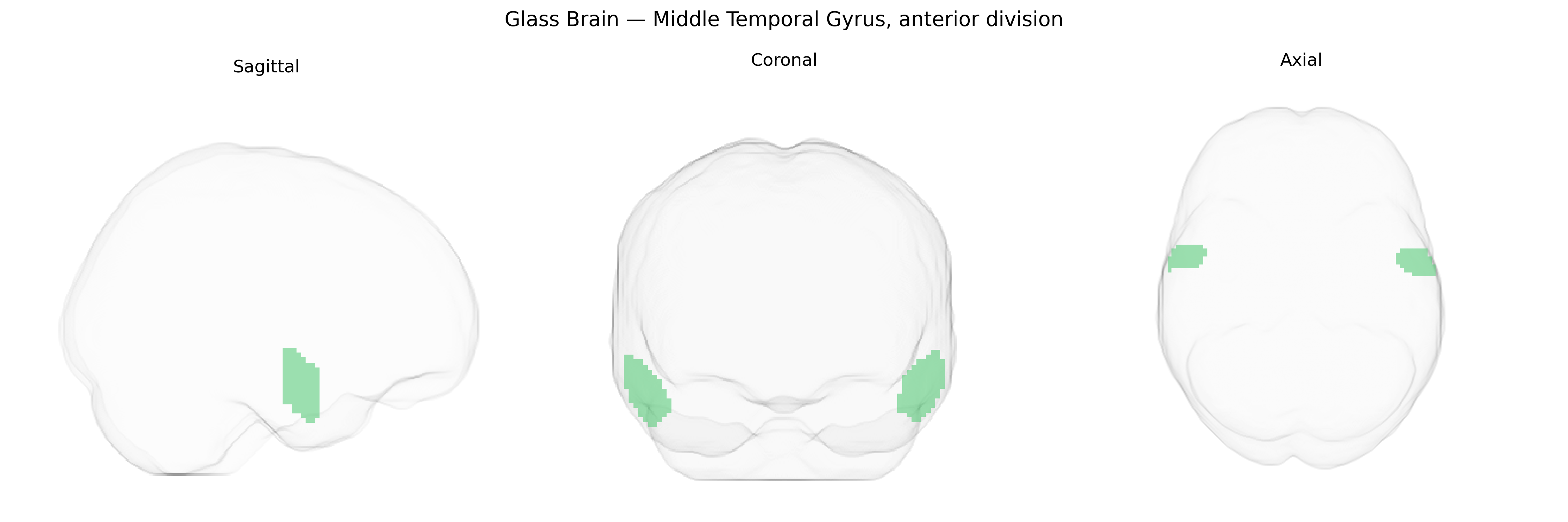
The bilateral Middle Temporal Gyrus, anterior division, as defined in the Harvard-Oxford cortical maximum probability atlas (threshold 25%, 2 mm), corresponds to the rostral portion of the middle temporal gyrus located on the lateral surface of the temporal lobe, extending anteriorly from approximately the level of the temporal pole and lying between the superior and inferior temporal gyri. This region is primarily associated with higher-order auditory and language processing, including semantic comprehension and integration of lexical information, and it contributes to multimodal association functions by linking auditory, visual, and conceptual representations. It receives inputs from primary and secondary auditory cortices as well as association areas in the frontal and parietal lobes, and projects to distributed language and semantic networks, including parts of the inferior frontal gyrus. Functionally and cytoarchitectonically, it is often grouped within the broader Middle Temporal Gyrus, a key component of the temporal lobe’s association cortex.
Genetic associations involving the bilateral anterior middle temporal gyrus (MTG) from the Harvard–Oxford atlas arise mainly from imaging–genetics and GWAS of cortical morphology and functional networks rather than region-specific candidate studies; large consortia such as ENIGMA and UK Biobank have shown that common variants near genes involved in neurodevelopment, synaptic function, and axon guidance (for example, variants in or near genes like WNT signaling components, cell-adhesion molecules, and glutamatergic synapse genes) are associated with cortical thickness and surface area measures that include the anterior MTG, though these effects are usually reported at the lobe or broader temporal subregion level rather than this specific atlas parcel. Functionally, the anterior MTG participates in semantic processing, language, and social cognition, and genetic risk for neuropsychiatric and neurodevelopmental disorders that impact these domains—such as schizophrenia, autism spectrum disorder, and primary progressive aphasia—has been statistically linked in imaging–genetics work to structural and functional alterations in anterior temporal regions, including the MTG, with polygenic risk scores for schizophrenia and autism showing associations with temporal lobe cortical morphology and connectivity. GWAS of language and cognitive traits have identified loci influencing temporal lobe structure and activation, and some Alzheimer’s disease and frontotemporal dementia risk variants are associated with atrophy patterns that encompass the anterior MTG, but the current literature rarely isolates the “bilateral Middle Temporal Gyrus, anterior division” as defined in the Harvard–Oxford cort maxprob thr25 2 mm atlas, so existing genetic findings should be interpreted as applying to overlapping or broader anterior temporal territories rather than this exact parcellation.
Overview generated by GPT-4o (2026).
Region ID: 11
Hemisphere: bilateral
Atlas: HarvardOxford cort maxprob thr25 2mm
Full Quality Version: Download MP4
Full Quality Version: Download MP4
Wali Sidiqyar*, Gaurav Rudravaram*, Elyssa M. McMaster, Trent M. Schwartz, Adam M. Saunders, Kurt G. Schilling, Bennett A. Landman "Introducing SPINS: A Shared Public Visualization Library of Neuroanatomical Structures." Medical Imaging with Deep Learning- short paper
This resource is licensed under CC0 1.0 Universal (Public Domain).