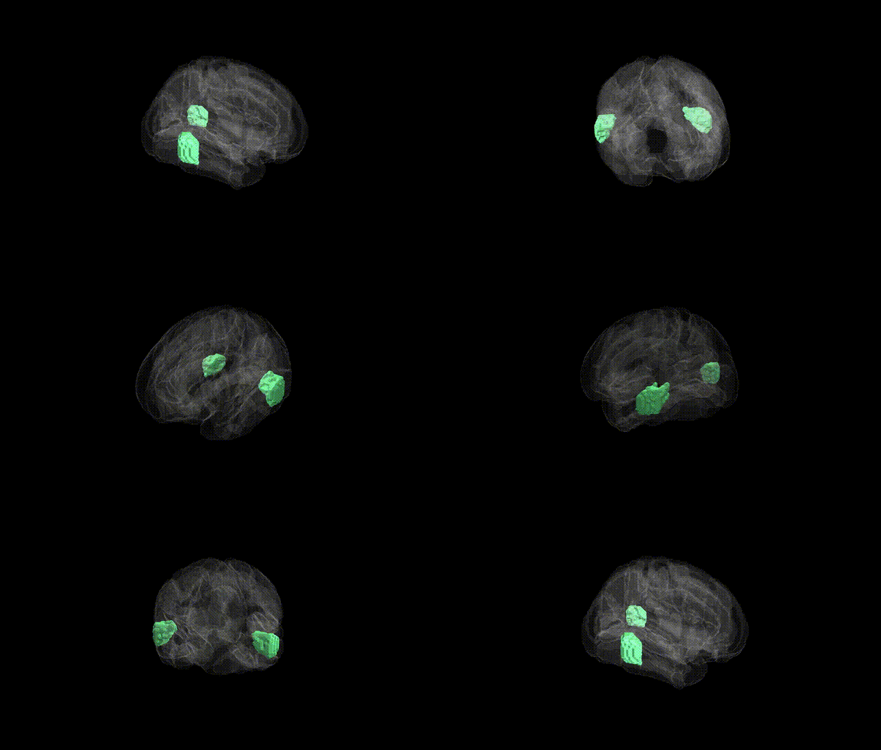
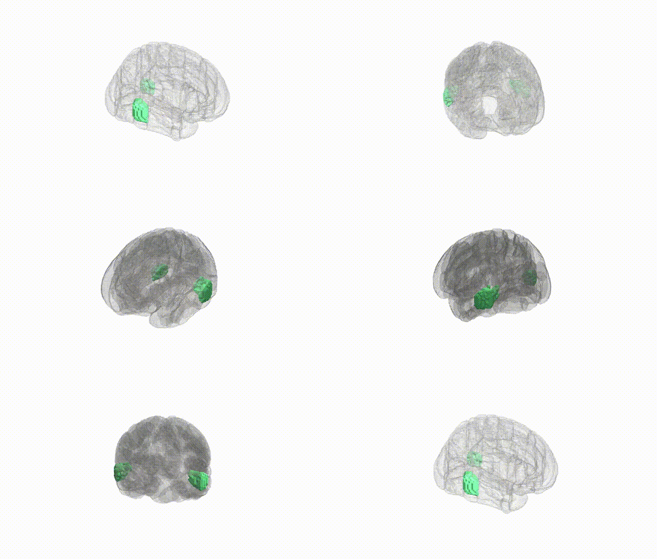
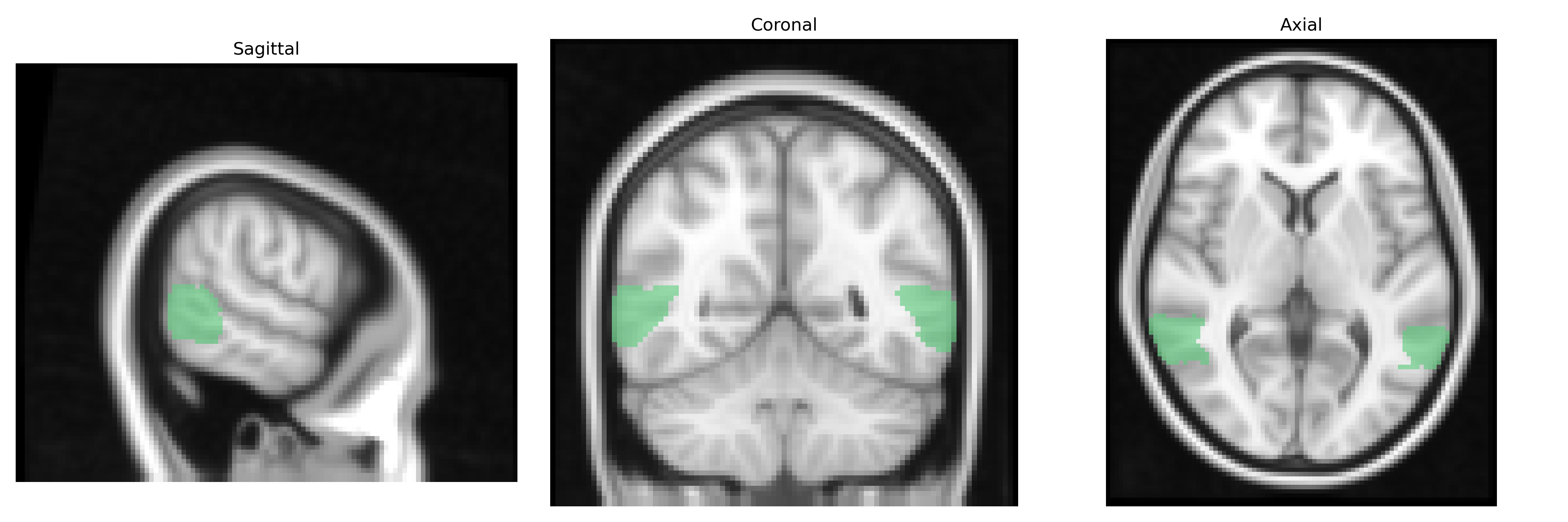
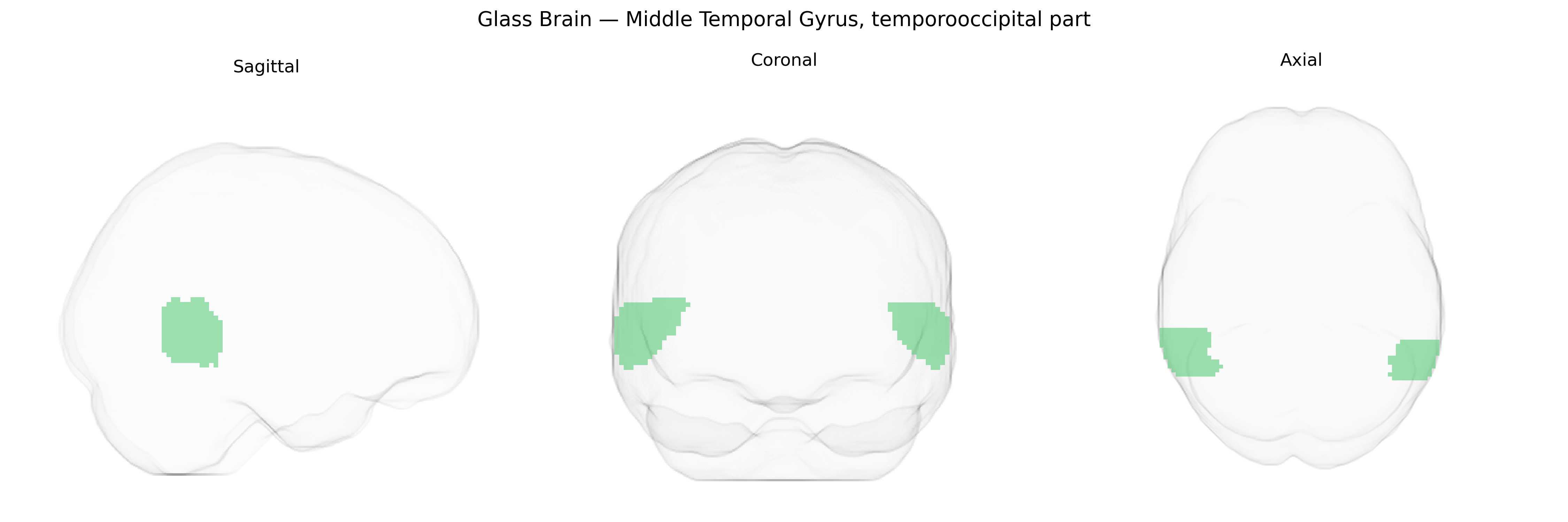
The bilateral Middle Temporal Gyrus, temporooccipital part, as defined in the Harvard–Oxford cortical maxprob thr25 2 mm atlas, corresponds to the posterior segment of the middle temporal gyrus located near the temporal–occipital junction of both hemispheres. This region lies along the lateral aspect of the temporal lobe and extends posteriorly toward the occipital lobe, forming part of the ventral visual processing stream. Functionally, it is implicated in high-level visual perception and object recognition, language and semantic processing, and the integration of auditory and visual information. It also participates in networks supporting reading and lexical access, and may contribute to multimodal association processes linking visual stimuli with conceptual or linguistic representations. There is no direct Wikipedia article for this exact subdivision; a closely related structure is the broader Middle temporal gyrus.
The bilateral middle temporal gyrus, temporooccipital part (posterior MTG/temporo‑occipital junction) from the Harvard–Oxford atlas has been implicated genetically mainly through large-scale imaging–genetics and GWAS meta-analyses of cortical structure and function rather than region-specific candidate-gene studies. Variants in genes influencing cortical surface area and thickness (for example in pathways involving WNT signaling, neuronal migration, and synaptic development such as TBR1, EOMES-related networks, and common neurodevelopmental loci identified by ENIGMA and UK Biobank imaging GWAS) show significant associations with temporal and occipital association cortex measures that include the posterior MTG. Polygenic architectures overlapping with educational attainment, general cognitive ability, and reading-related traits have been linked to structural variation in posterior temporal regions that encompass this area, consistent with its role in semantic processing, language, and visual word recognition. Shared genetic risk across schizophrenia, autism spectrum disorder, and major depressive disorder has been associated with altered structure or connectivity in lateral temporal cortices, including the temporo‑occipital MTG, in imaging–genetic and PRS (polygenic risk score) studies, although no single locus has been uniquely tied to this subregion. GWAS of functional networks and resting-state connectivity also implicate common variants affecting temporo‑occipital association networks involved in higher-order visual, language, and default-mode–related processing, suggesting that genetic influences on this region are distributed and pleiotropic rather than specific to a single disease or trait.
Overview generated by GPT-4o (2026).
Region ID: 13
Hemisphere: bilateral
Atlas: HarvardOxford cort maxprob thr25 2mm
Full Quality Version: Download MP4
Full Quality Version: Download MP4
Wali Sidiqyar*, Gaurav Rudravaram*, Elyssa M. McMaster, Trent M. Schwartz, Adam M. Saunders, Kurt G. Schilling, Bennett A. Landman "Introducing SPINS: A Shared Public Visualization Library of Neuroanatomical Structures." Medical Imaging with Deep Learning- short paper
This resource is licensed under CC0 1.0 Universal (Public Domain).