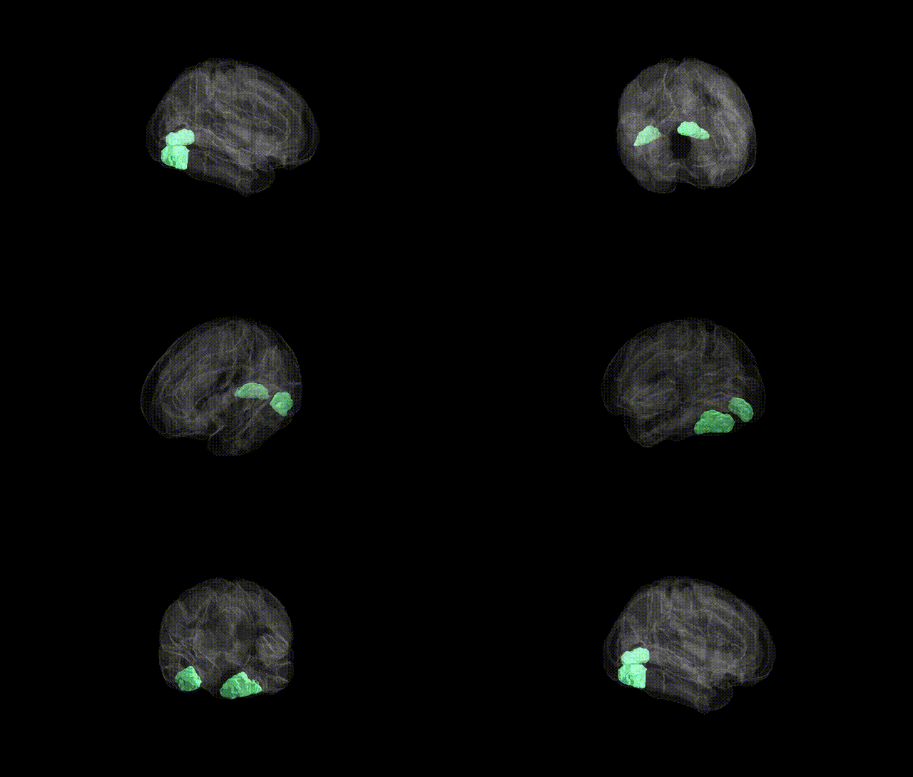
The bilateral Occipital Fusiform Gyrus, as defined in the Harvard-Oxford cortical atlas, comprises fusiform cortex located in the occipital lobe on both hemispheres, lying ventral to the lingual gyrus and posterior to the temporal fusiform regions along the ventral visual pathway. This region is primarily associated with high-level visual processing, including aspects of object and form recognition, and contributes to the transformation of early visual input into more complex representations. It forms part of the broader fusiform gyrus, which is a key component of the ventral temporal–occipital cortex involved in visual perception and category-specific processing. There is no direct Wikipedia article for “Occipital Fusiform Gyrus”; a closely related and encompassing structure is the Fusiform gyrus.
The occipital fusiform gyrus, corresponding largely to ventral occipital and posterior fusiform visual association cortex in the Harvard-Oxford atlas, has been implicated in several genetic and GWAS-based associations, though most findings are indirect via imaging genetics rather than region-specific GWAS. Variants in genes influencing cortical surface area and thickness (such as those near HMGA2, KIAA0586, and multiple loci in large ENIGMA consortium studies) have been associated with structural differences in ventral occipital–fusiform regions, particularly in visual recognition networks. Polygenic risk scores for schizophrenia, bipolar disorder, and major depressive disorder show associations with altered fusiform and occipital cortex volumes and functional connectivity, suggesting genetically driven vulnerability in visual and socio-cognitive processing circuits. GWAS of face recognition and prosopagnosia have pointed to heritable variation in fusiform face area function, with some studies implicating loci in neurotransmission and synaptic plasticity pathways (e.g., glutamatergic and GABAergic genes) that modulate activity in fusiform/occipital visual regions. Autism spectrum disorder and dyslexia imaging-genetic studies have linked common variants (including in CNTNAP2, DCDC2, and related neurodevelopmental genes) to atypical activation and structural differences in occipito-temporal cortex encompassing the occipital fusiform gyrus during face, word, and object processing. Additionally, GWAS of general cognitive ability and educational attainment demonstrate distributed cortical effects that include occipito-temporal areas involved in high-level visual processing, indicating that polygenic influences on cognition and neurodevelopment partially manifest through variability in this bilateral region.
Overview generated by GPT-4o (2026).
Region ID: 40
Hemisphere: bilateral
Atlas: HarvardOxford cort maxprob thr25 2mm
Full Quality Version: Download MP4
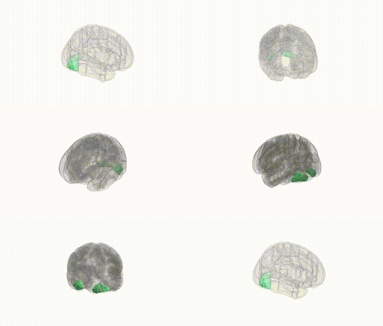
Full Quality Version: Download MP4
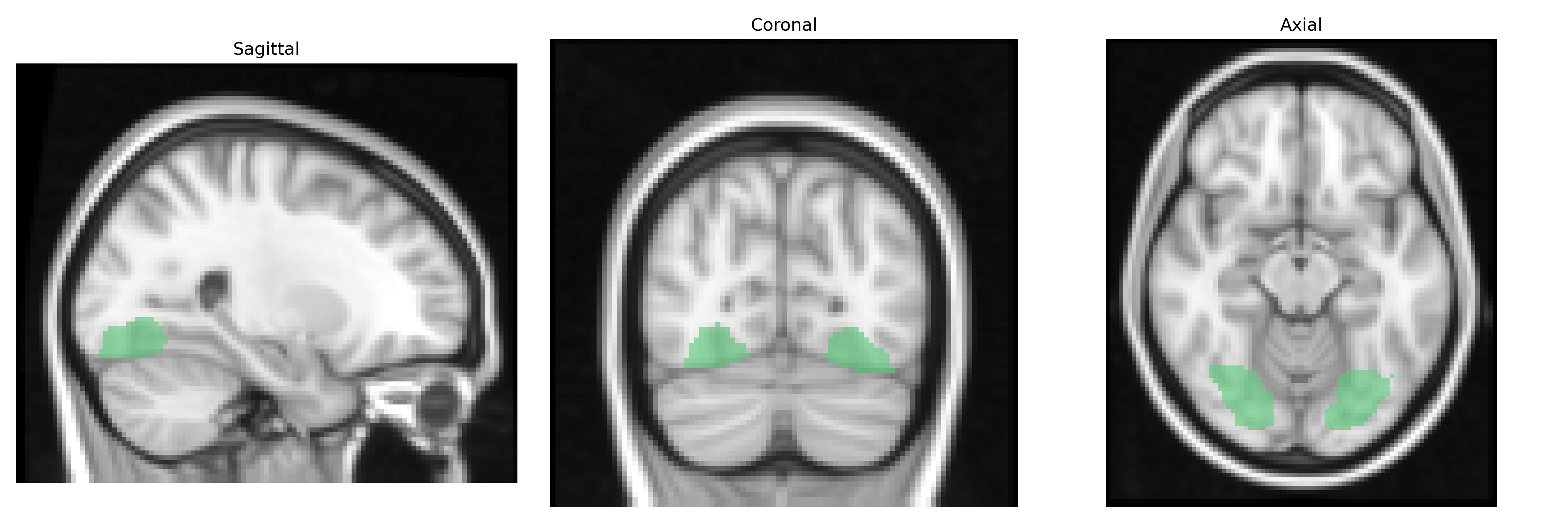
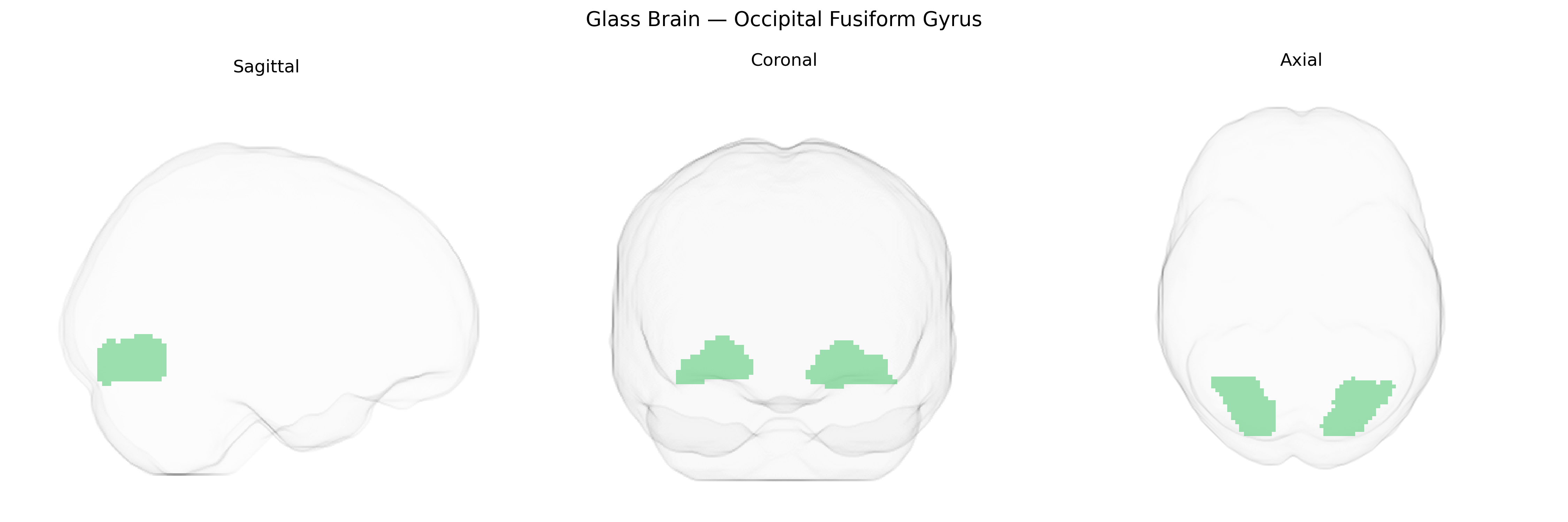
Wali Sidiqyar*, Gaurav Rudravaram*, Elyssa M. McMaster, Trent M. Schwartz, Adam M. Saunders, Kurt G. Schilling, Bennett A. Landman "Introducing SPINS: A Shared Public Visualization Library of Neuroanatomical Structures." Medical Imaging with Deep Learning- short paper
This resource is licensed under CC0 1.0 Universal (Public Domain).