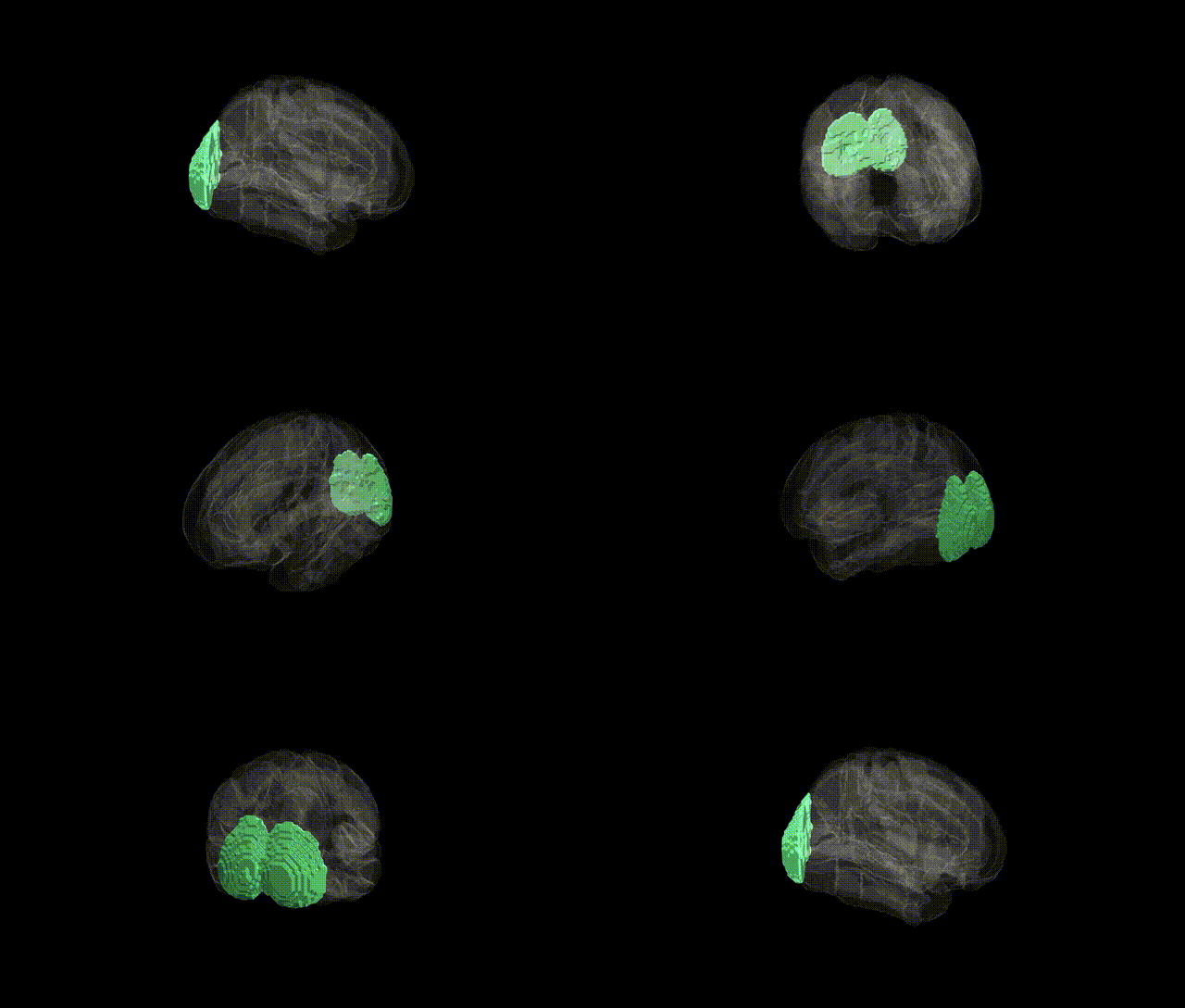
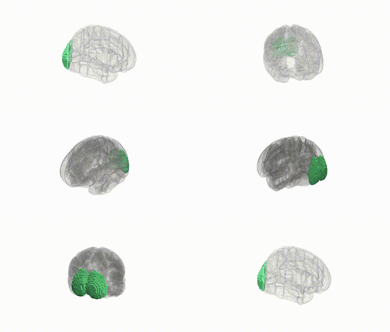
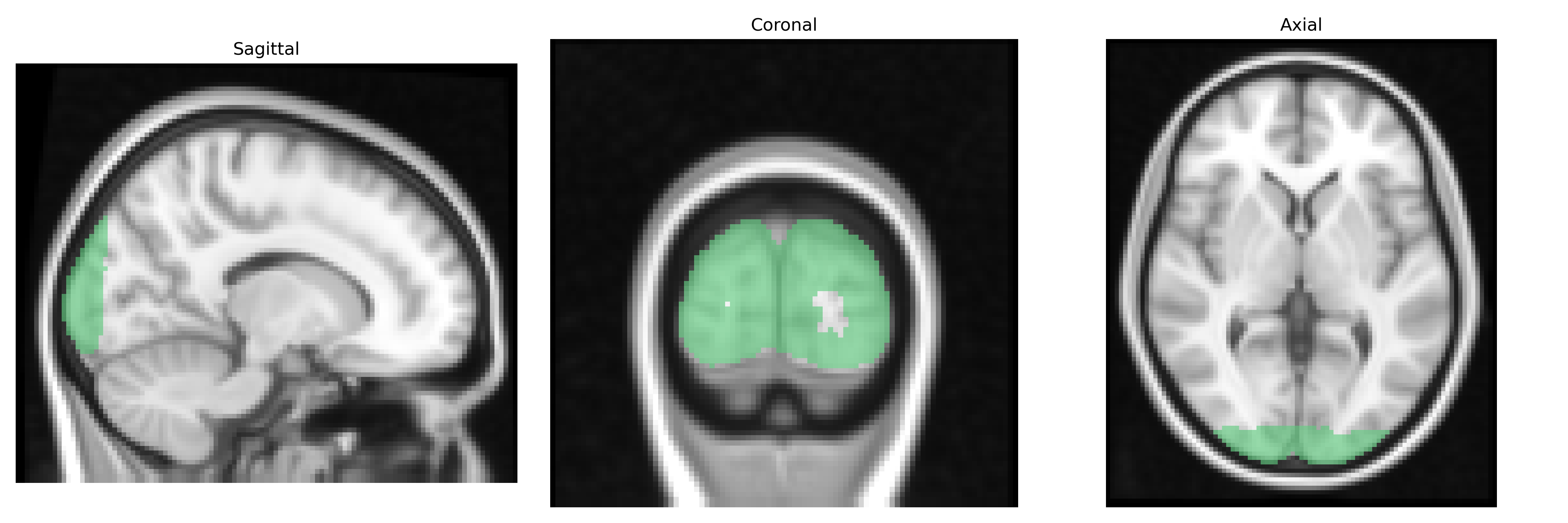
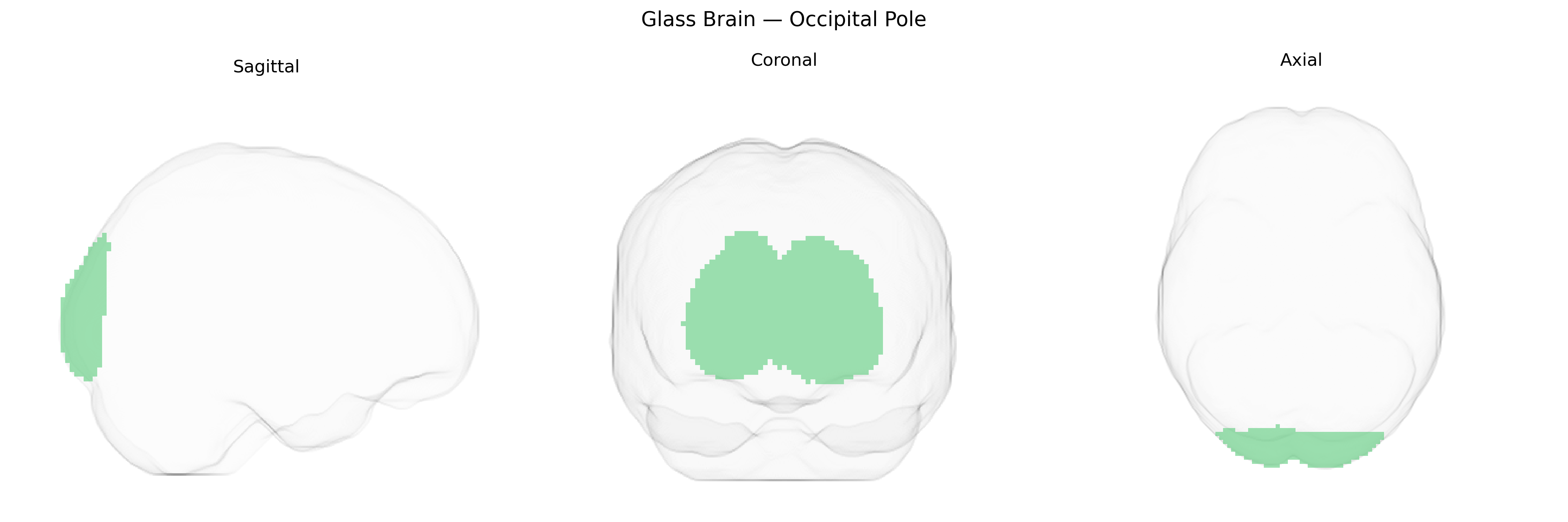
The bilateral Occipital Pole, as defined in the Harvard–Oxford cortical atlas, corresponds to the most posterior part of the occipital lobes, encompassing primary and adjacent early visual cortical areas involved in the initial processing of visual information. This region contains portions of the striate and extrastriate cortex that receive direct thalamic input from the lateral geniculate nucleus and participate in encoding basic visual features such as luminance, contrast, orientation, motion, and spatial frequency. Functionally, the Occipital Pole acts as an early-stage relay and transformation hub for visual signals that are subsequently distributed along dorsal and ventral visual streams to support visually guided actions and object recognition. Anatomically, it lies posterior to the parieto-occipital sulcus and above and below the calcarine sulcus, with extensive interhemispheric connectivity via the splenium of the corpus callosum. There is no direct Wikipedia article for the “Occipital Pole” as an atlas-defined region; a closely related structure is the Occipital lobe.
The bilateral occipital pole, corresponding to the primary and early visual cortex at the posterior extreme of the occipital lobes, shows genetic associations largely through studies of cortical thickness, surface area, and visual processing traits rather than direct disease-specific signals. Twin and imaging-genetics studies indicate high heritability of occipital pole morphology, with variants in genes involved in neurodevelopment and synaptic function (e.g., regulators of axon guidance, neuronal migration, and glutamatergic signaling) contributing to structural variation, though specific loci are often shared across occipital and broader cortical regions. GWAS of cortical measures have identified common variants near genes such as PAX6, TBR1, and other transcription factors important for occipital and visual cortex development, with some signals overlapping with polygenic architectures of general cognitive ability and educational attainment. Occipital pole measures have also appeared in imaging-GWAS of psychiatric and neurodevelopmental disorders, including schizophrenia, major depression, and autism spectrum conditions, where risk variants influence occipital cortical thickness or volume as part of distributed brain-wide effects, although these associations are typically modest and not specific to this region. Additionally, genetic studies of visual traits (e.g., visual acuity, susceptibility to visual hallucinations, migraine with aura) and neurological disorders involving visual symptoms have implicated occipital regions, including the pole, through functional imaging and polygenic risk analyses, but precise gene–region links remain diffuse and largely reflect shared genetic influences on cortical development and connectivity rather than occipital pole–exclusive effects.
Overview generated by GPT-4o (2026).
Region ID: 48
Hemisphere: bilateral
Atlas: HarvardOxford cort maxprob thr25 2mm
Full Quality Version: Download MP4
Full Quality Version: Download MP4
Wali Sidiqyar*, Gaurav Rudravaram*, Elyssa M. McMaster, Trent M. Schwartz, Adam M. Saunders, Kurt G. Schilling, Bennett A. Landman "Introducing SPINS: A Shared Public Visualization Library of Neuroanatomical Structures." Medical Imaging with Deep Learning- short paper
This resource is licensed under CC0 1.0 Universal (Public Domain).