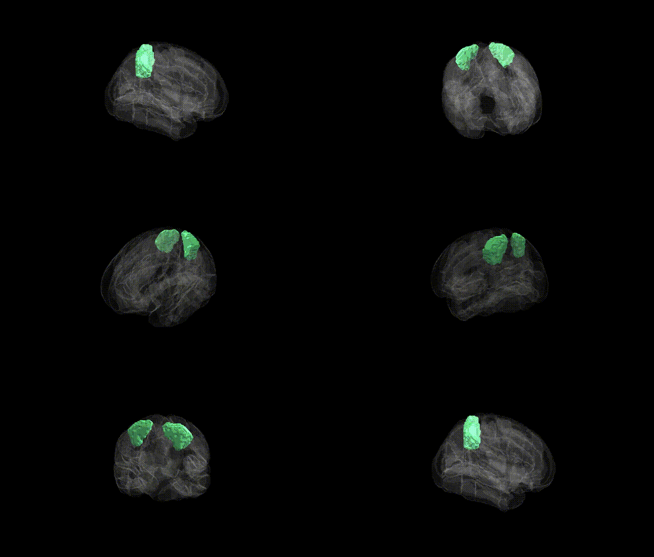
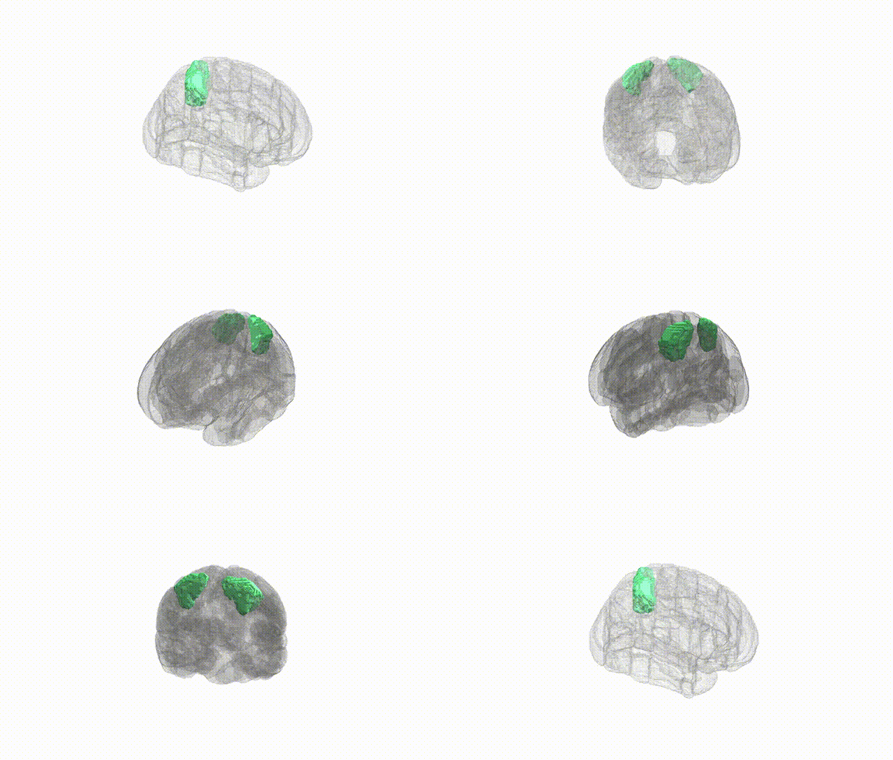
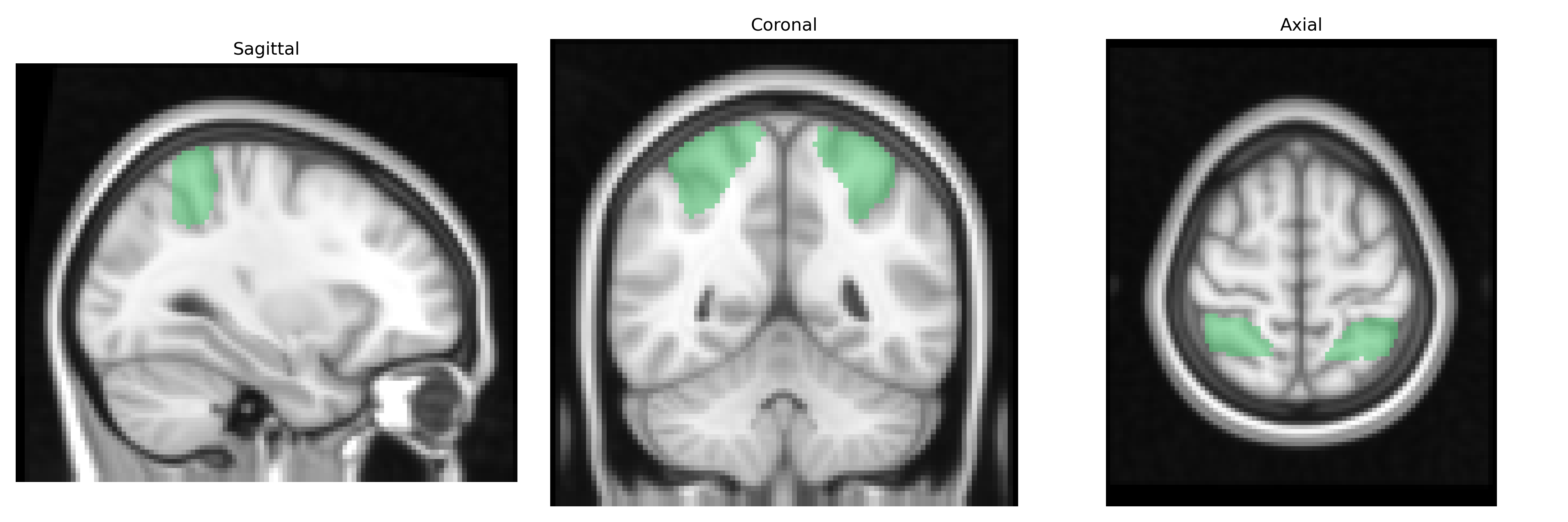
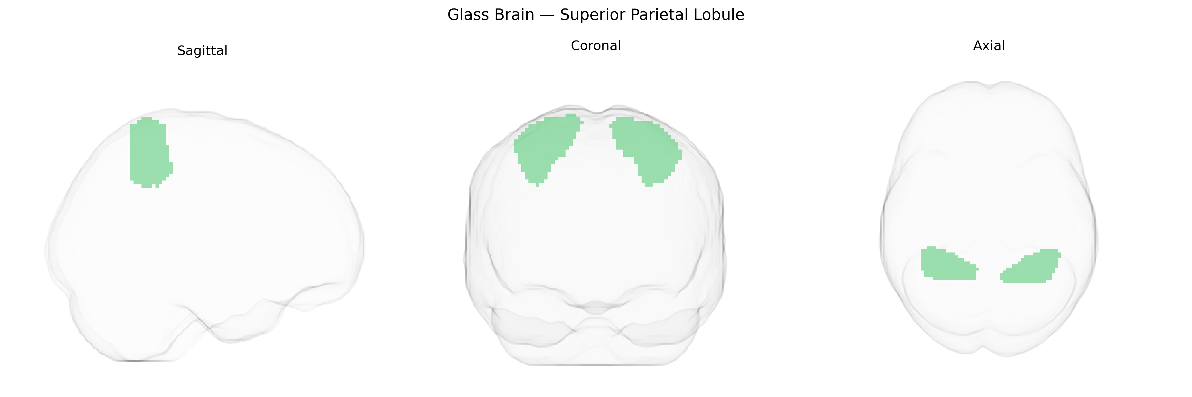
The bilateral Superior Parietal Lobule (SPL), as defined in the Harvard-Oxford cortical maxprob thr25 2mm atlas, comprises dorsal parietal cortex lying posterior to the postcentral gyrus and superior to the intraparietal sulcus in both hemispheres, typically corresponding to Brodmann areas 7 and parts of 5. This region is heavily involved in visuospatial processing, multisensory integration, and the coordination of attention and sensorimotor transformations necessary for reaching, grasping, and complex hand–eye coordination. The SPL contributes to spatial working memory, body-centered reference frames, and the integration of proprioceptive, visual, and tactile information, and is considered a key node in dorsal attention and fronto-parietal control networks. Functional and lesion studies implicate this region in higher-order aspects of perception and action, including mental rotation, spatial navigation, and the maintenance of spatial representations that support goal-directed behavior.
Superior parietal lobule
The bilateral Superior Parietal Lobule (SPL), as defined in the HarvardOxford cortical atlas, has been implicated in genetic studies primarily through imaging genetics and GWAS of brain structure and cognition rather than direct region-specific association scans. Variants in genes involved in synaptic function and neurodevelopment—such as those in the BDNF, COMT, and CNTNAP2 pathways—have been associated with individual differences in parietal cortical thickness, surface area, or activation in tasks requiring visuospatial attention, working memory, and sensorimotor integration, functions strongly linked to SPL. Large-scale GWAS of cortical morphology (e.g., ENIGMA consortium and UK Biobank studies) have identified multiple loci (including regions near microtubule, cell adhesion, and chromatin-remodeling genes) that influence parietal lobe measures, though these findings generally aggregate across parietal subdivisions and do not isolate SPL specifically. SPL structure and connectivity show heritability and have been indirectly associated with genetic risk for neurodevelopmental and psychiatric conditions, including schizophrenia, autism spectrum disorder, attention-deficit/hyperactivity disorder, and dyslexia, where risk variants often affect distributed frontoparietal networks. In addition, polygenic scores for general cognitive ability and educational attainment correlate with parietal cortical metrics encompassing SPL, supporting a genetically mediated role of this region in higher-order cognition, while GWAS on traits such as mathematical ability and visuospatial skills have reported parietal-involving networks but without SPL-specific loci. Overall, genetic associations with SPL are currently best characterized at the level of parietal or frontoparietal networks and cortical morphology, with few findings uniquely targeting the bilateral SPL region as defined in the HarvardOxford atlas.
Overview generated by GPT-4o (2026).
Region ID: 18
Hemisphere: bilateral
Atlas: HarvardOxford cort maxprob thr25 2mm
Full Quality Version: Download MP4
Full Quality Version: Download MP4
Wali Sidiqyar*, Gaurav Rudravaram*, Elyssa M. McMaster, Trent M. Schwartz, Adam M. Saunders, Kurt G. Schilling, Bennett A. Landman "Introducing SPINS: A Shared Public Visualization Library of Neuroanatomical Structures." Medical Imaging with Deep Learning- short paper
This resource is licensed under CC0 1.0 Universal (Public Domain).