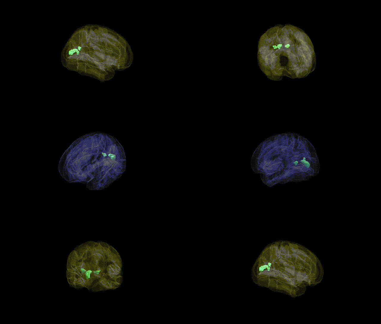
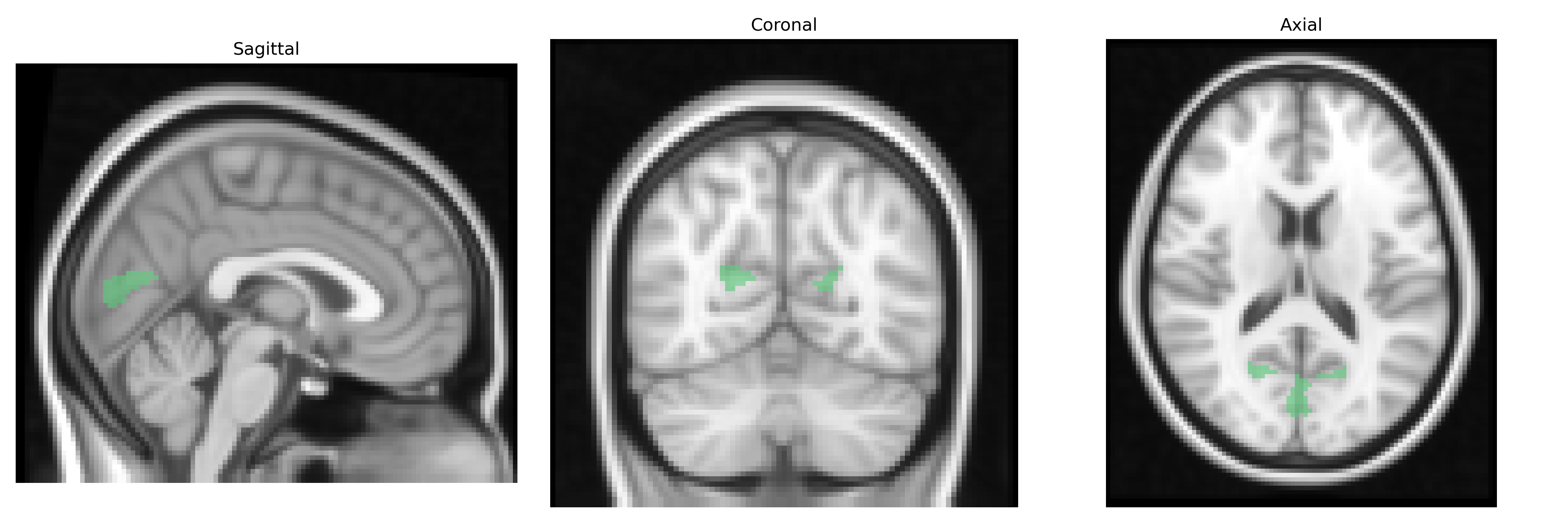
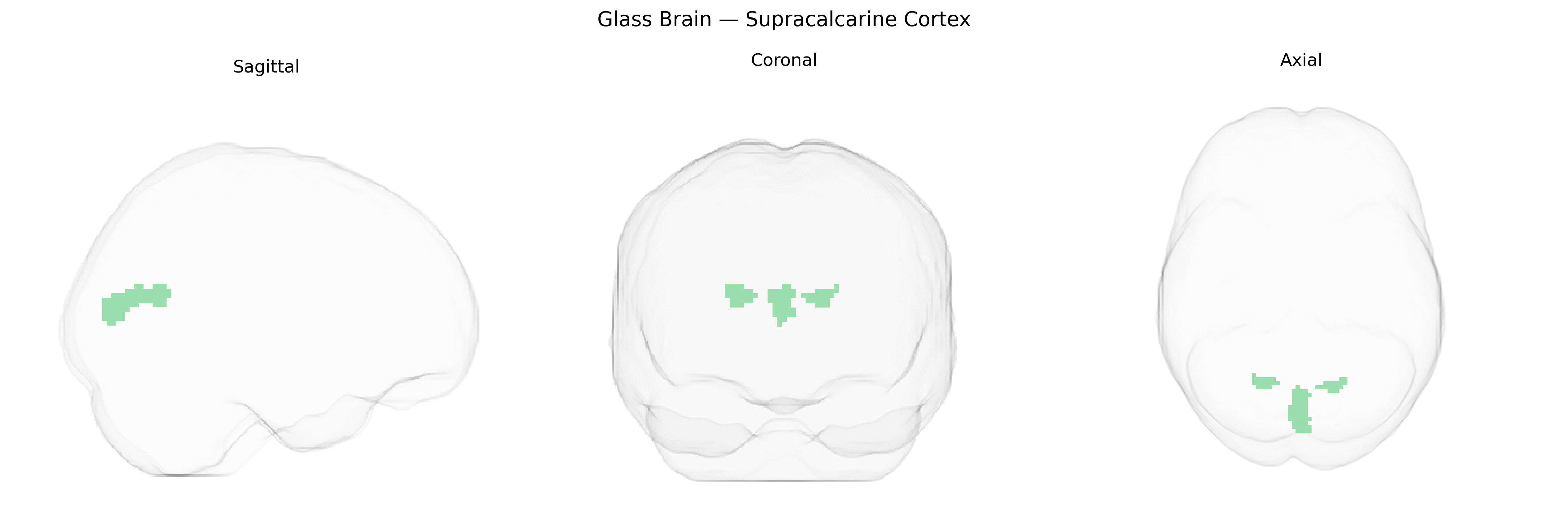
The bilateral supracalcarine cortex, as defined in the Harvard–Oxford Cortical Structural Atlas, refers to a portion of occipital cortex located superior to the calcarine sulcus and closely associated with the primary visual cortex (V1) and adjacent early visual areas. This region participates in initial stages of visual processing, including the analysis of basic features such as orientation, contrast, and spatial frequency, and contributes to the retinotopic mapping of the visual field. Structurally, it lies within the medial occipital lobe, bordering the calcarine cortex and connecting with other visual areas through dense intracortical projections. There is no direct Wikipedia article for the supracalcarine cortex; a related and encompassing structure is the Primary visual cortex.
The bilateral supracalcarine cortex, encompassing primary and pericalcarine visual areas, shows genetic associations largely through GWAS of brain structure and function rather than direct region-specific studies; SNP-based heritability analyses from large neuroimaging consortia (e.g., ENIGMA, UK Biobank) indicate moderate to high heritability of occipital cortical thickness and surface area, with loci near genes such as HMGA2, IGF1, and MIR148A implicated in global and posterior cortical morphology that include the supracalcarine region. Polygenic risk for neuropsychiatric disorders—particularly schizophrenia, bipolar disorder, and major depression—has been associated with altered occipital and calcarine cortical thickness or volume, suggesting shared genetic architecture influencing both psychiatric liability and visual cortex structure. Genome-wide studies of visual processing traits and resting-state networks have linked variants in synaptic and neurodevelopmental genes (e.g., NRXN1, GRIN2B, BDNF-related pathways) to functional connectivity and activation patterns in early visual cortex, including calcarine and supracalcarine areas, while rare variant and copy-number studies in developmental disorders (autism spectrum conditions, intellectual disability, and congenital visual impairment) frequently implicate genes involved in axon guidance and cortical patterning that affect occipital lobe organization. Overall, genetic evidence supports a multifactorial architecture in which common polymorphisms and rare variants in neurodevelopmental, synaptic, and growth-regulating genes contribute to interindividual differences in supracalcarine cortex anatomy and function and to its involvement in neurodevelopmental and psychiatric disorders.
Overview generated by GPT-4o (2026).
Region ID: 47
Hemisphere: bilateral
Atlas: HarvardOxford cort maxprob thr25 2mm
Full Quality Version: Download MP4
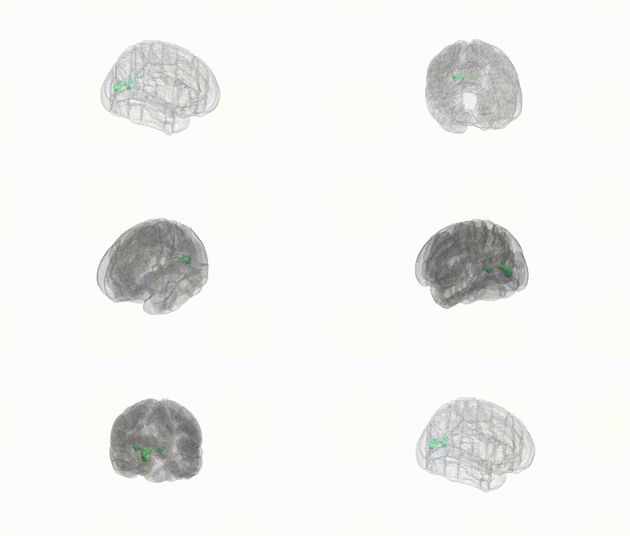
Full Quality Version: Download MP4
Wali Sidiqyar*, Gaurav Rudravaram*, Elyssa M. McMaster, Trent M. Schwartz, Adam M. Saunders, Kurt G. Schilling, Bennett A. Landman "Introducing SPINS: A Shared Public Visualization Library of Neuroanatomical Structures." Medical Imaging with Deep Learning- short paper
This resource is licensed under CC0 1.0 Universal (Public Domain).