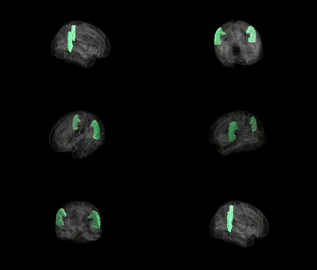
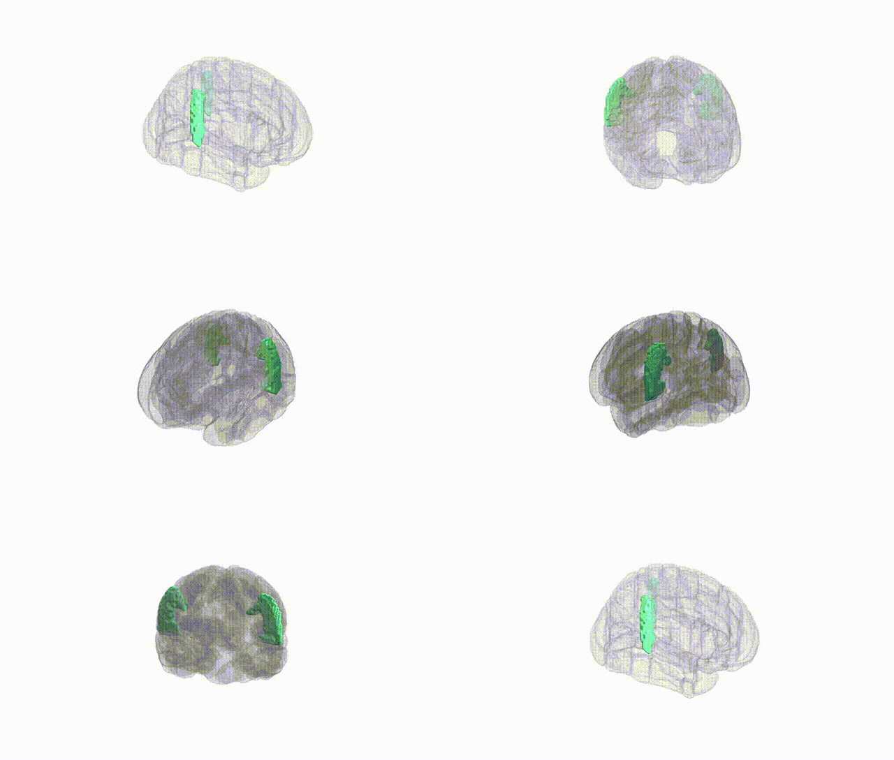
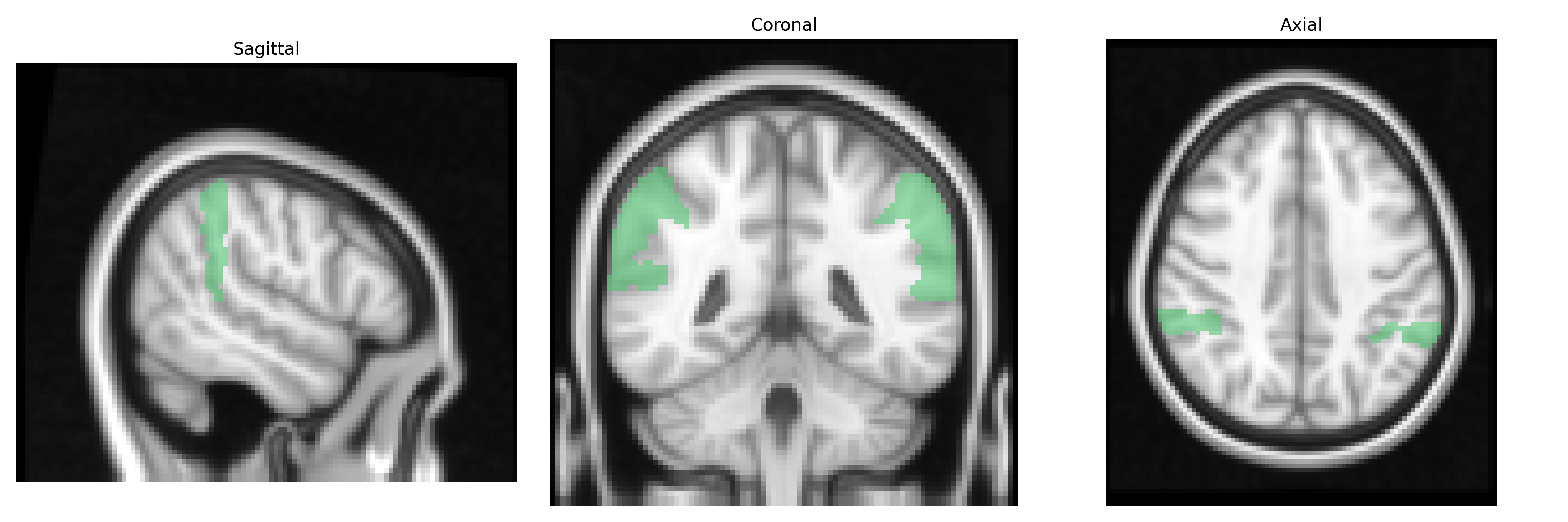
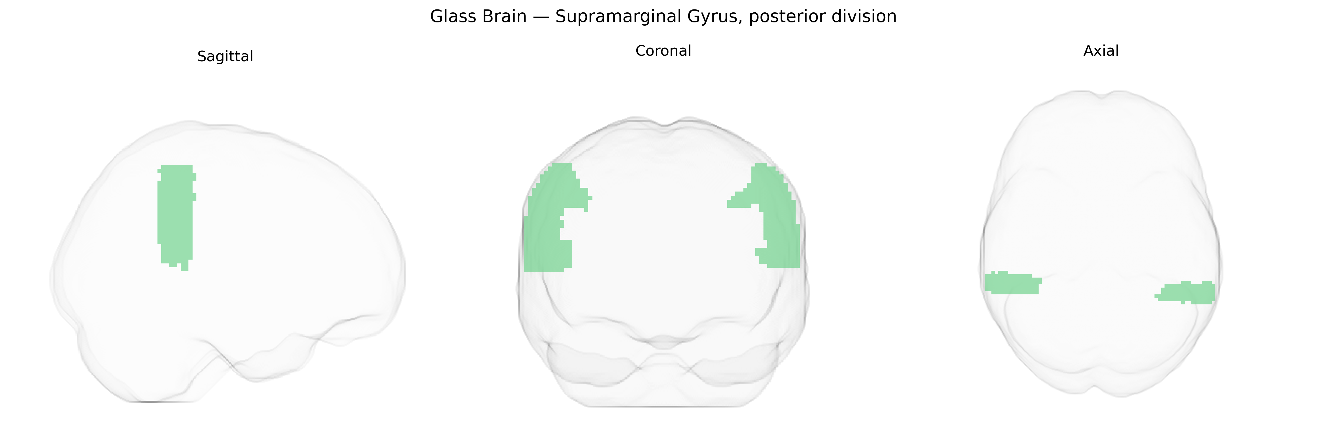
The bilateral Supramarginal Gyrus, posterior division, as defined in the Harvard-Oxford cortical atlas, corresponds to the caudal portion of the supramarginal gyrus wrapping around the posterior end of the lateral (Sylvian) fissure and forming part of the inferior parietal lobule. It lies dorsal to the superior temporal gyrus and anterior to the angular gyrus, integrating multimodal sensory inputs—particularly auditory, somatosensory, and visual information—and is implicated in phonological processing, language comprehension, and aspects of social cognition such as empathy and perspective-taking, with hemispheric specializations (left more often associated with language, right with spatial and socio-emotional functions). Cytoarchitectonically, it is considered part of the inferior parietal cortex and shows strong connectivity with temporal, frontal, and other parietal association areas, supporting its role in higher-order integration of perception, action, and language. Supramarginal gyrus
The bilateral supramarginal gyrus, posterior division, as defined in the Harvard–Oxford atlas, has been implicated in genetic studies primarily through imaging-genetics and GWAS of cortical structure and cognition rather than direct region-specific GWAS hits. Large-scale MRI-based GWAS (e.g., ENIGMA and UK Biobank) have identified common variants in genes involved in neurodevelopment, synaptic function, and cell adhesion (such as HMGA2, MIR588, and IGF1 pathway genes) associated with supramarginal or inferior parietal cortical thickness and surface area, with heritability estimates in the moderate range. Polygenic risk for schizophrenia, major depressive disorder, and bipolar disorder has been linked to altered morphology of inferior parietal and supramarginal regions, with some studies reporting genotype–phenotype correlations involving DISC1-related pathways, CACNA1C, and other psychiatric risk loci that influence parietal cortex structure and connectivity. GWAS of language, reading ability, and dyslexia have connected risk variants (e.g., in KIAA0319, DCDC2, and ROBO1) to structural and functional alterations in temporo-parietal regions encompassing the posterior supramarginal gyrus, consistent with its role in phonological processing and verbal working memory. Additional associations include links between APOE and parietal atrophy patterns in Alzheimer’s disease, as well as polygenic scores for general intelligence and educational attainment correlating with supramarginal gyrus morphology and activation, although these effects are generally distributed across a broader fronto-parietal network rather than specific to this subregion.
Overview generated by GPT-4o (2026).
Region ID: 20
Hemisphere: bilateral
Atlas: HarvardOxford cort maxprob thr25 2mm
Full Quality Version: Download MP4
Full Quality Version: Download MP4
Wali Sidiqyar*, Gaurav Rudravaram*, Elyssa M. McMaster, Trent M. Schwartz, Adam M. Saunders, Kurt G. Schilling, Bennett A. Landman "Introducing SPINS: A Shared Public Visualization Library of Neuroanatomical Structures." Medical Imaging with Deep Learning- short paper
This resource is licensed under CC0 1.0 Universal (Public Domain).