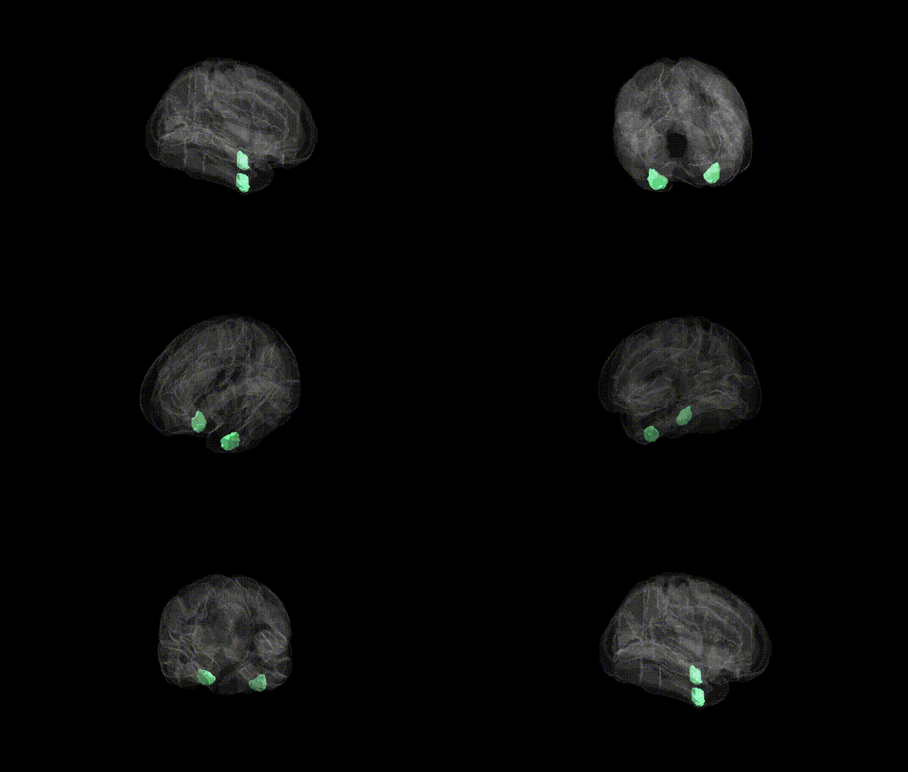
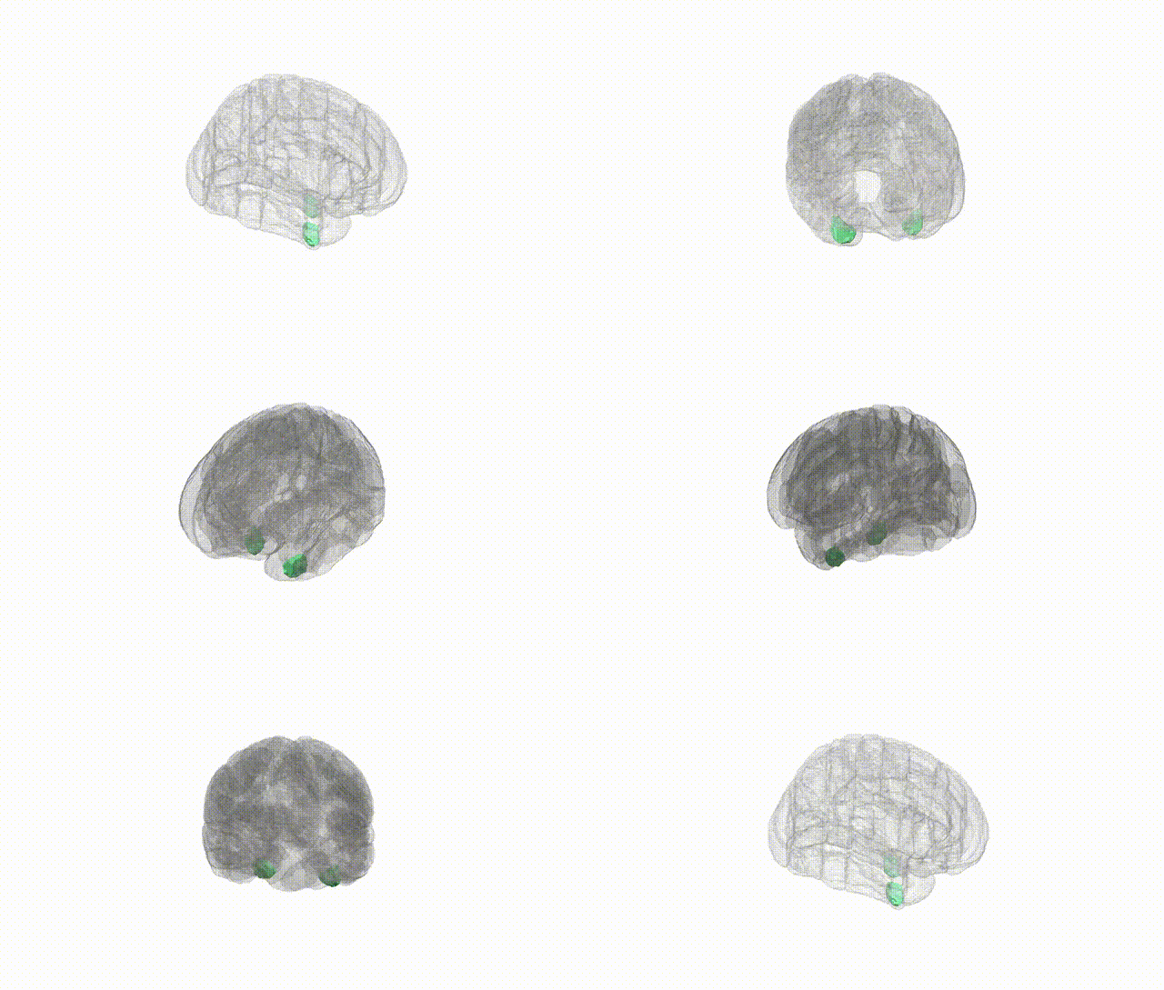
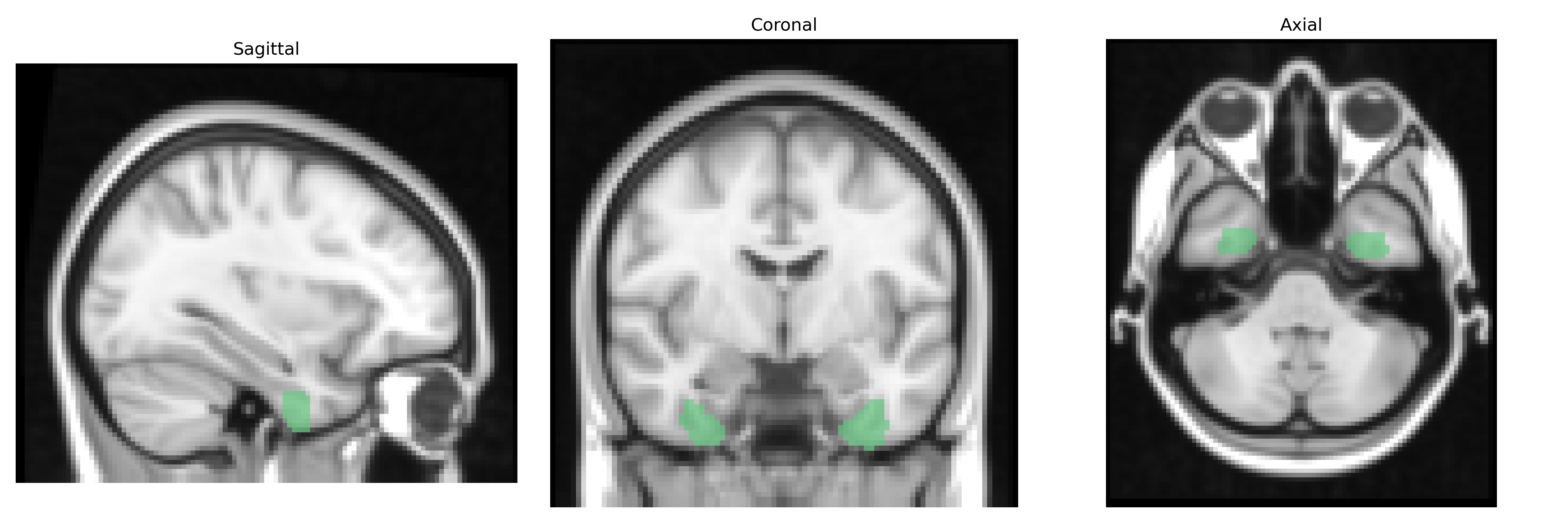
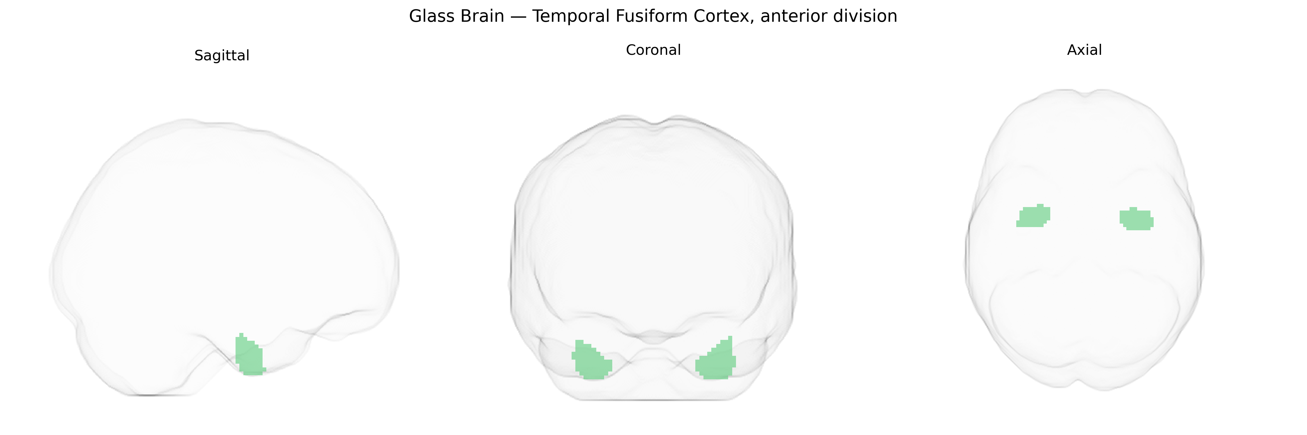
The bilateral Temporal Fusiform Cortex, anterior division (from the Harvard-Oxford Cortical Structural Atlas, maxprob thr25, 2 mm) corresponds to the anterior portion of the fusiform gyrus within the temporal lobe, located on the ventral surface between the inferior temporal gyrus laterally and the parahippocampal gyrus medially. This region is implicated in high-level visual processing, including aspects of object recognition, face and body perception, and integration of complex visual features with semantic information. Cytoarchitectonically, it forms part of the ventral visual stream and is interconnected with occipitotemporal, parietal, and limbic structures, supporting multimodal processing and memory-related functions. There is no direct Wikipedia article for “Temporal Fusiform Cortex, anterior division”; a closely related structure is the fusiform gyrus: Fusiform gyrus.
The bilateral Temporal Fusiform Cortex, anterior division, as defined in the Harvard–Oxford cortical atlas, overlaps closely with anterior fusiform and adjacent ventral temporal regions implicated in high-level visual processing, semantic memory, and social cognition, and genetic associations for this region are mostly inferred via broader fusiform/ventral temporal measures rather than this precise parcel. Large-scale imaging genetics studies (e.g., ENIGMA and UK Biobank analyses) have shown that cortical thickness and surface area in fusiform and anterior temporal regions are heritable and influenced by common variants in pathways related to neurodevelopment, synaptic signaling, and cell adhesion (including genes such as MAPT, BDNF, and several HLA and cadherin/axon guidance loci), although findings vary across studies and are rarely specific to the anterior fusiform subdivision. GWAS of face recognition and face perception abilities, as well as prosopagnosia risk, have implicated variants near genes involved in neural connectivity and plasticity, supporting a genetic contribution to functional specialization of fusiform regions, which include the anterior fusiform in extended face-processing networks. In neuropsychiatric and neurodevelopmental disorders, genetic risk for autism spectrum disorder, schizophrenia, and major depression has been associated with structural and functional alterations in fusiform and anterior temporal cortices, and polygenic risk scores for these conditions correlate with patterns of cortical thickness and connectivity in ventral temporal regions, though these effects are typically distributed and not localized uniquely to the Temporal Fusiform Cortex, anterior division. Overall, current genetic and GWAS evidence supports moderate heritability and polygenic influences on the morphology and function of fusiform/anterior temporal cortices, with links to social cognition, face processing, semantic networks, and vulnerability to neuropsychiatric and neurodevelopmental disorders, but region-specific genetic associations for this exact atlas parcel remain limited.
Overview generated by GPT-4o (2026).
Region ID: 37
Hemisphere: bilateral
Atlas: HarvardOxford cort maxprob thr25 2mm
Full Quality Version: Download MP4
Full Quality Version: Download MP4
Wali Sidiqyar*, Gaurav Rudravaram*, Elyssa M. McMaster, Trent M. Schwartz, Adam M. Saunders, Kurt G. Schilling, Bennett A. Landman "Introducing SPINS: A Shared Public Visualization Library of Neuroanatomical Structures." Medical Imaging with Deep Learning- short paper
This resource is licensed under CC0 1.0 Universal (Public Domain).