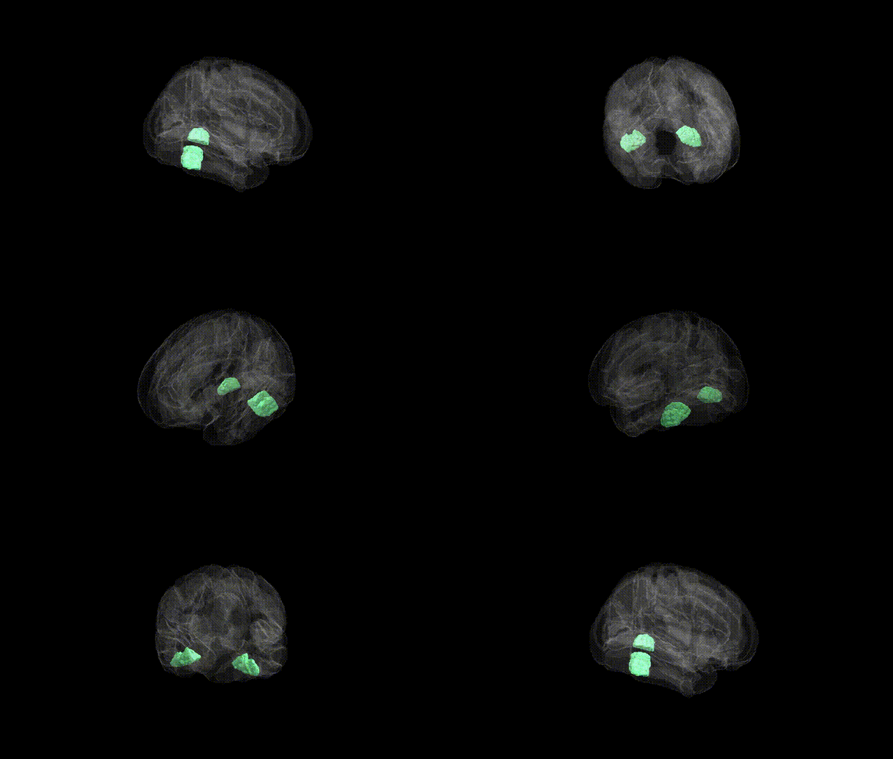
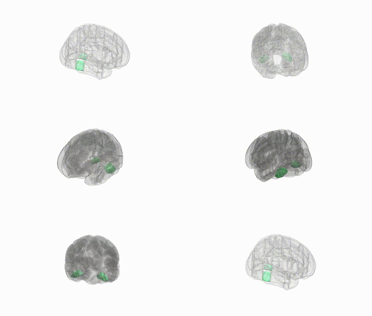
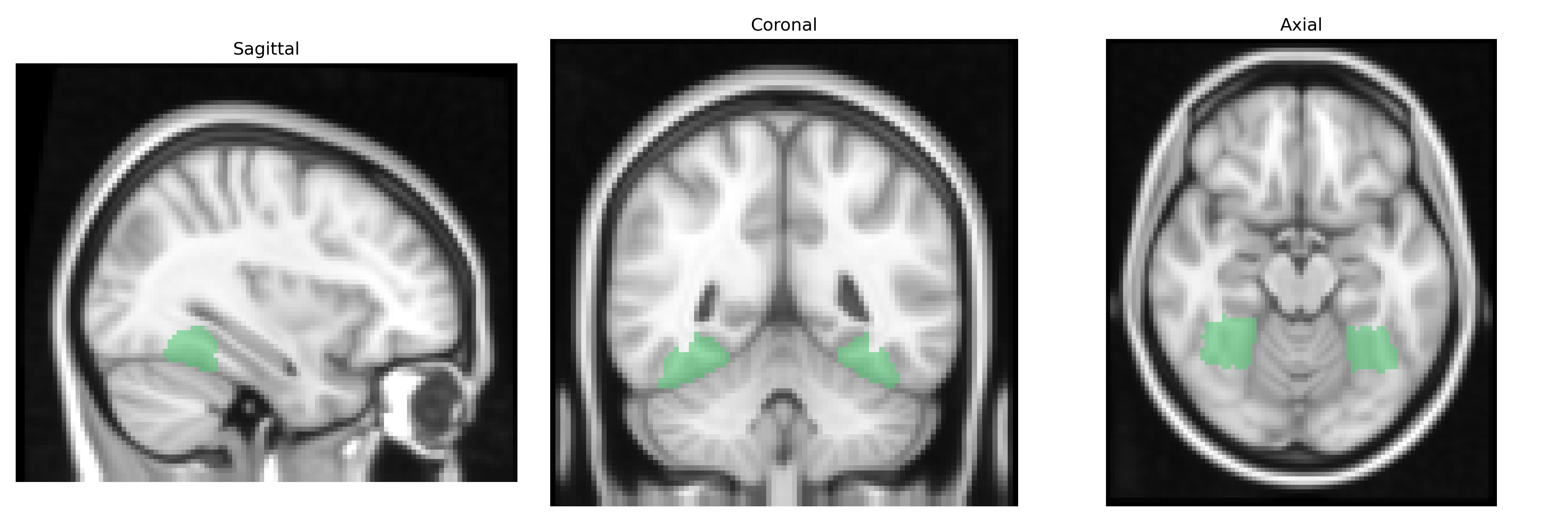
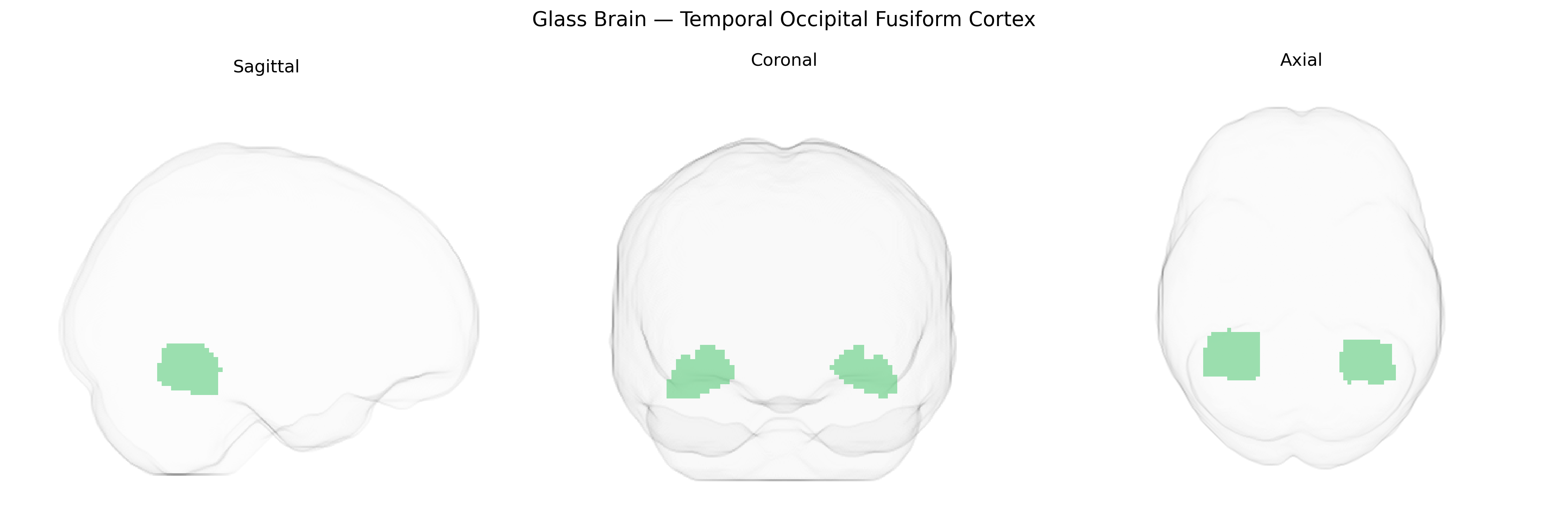
The bilateral Temporal Occipital Fusiform Cortex, as defined in the Harvard-Oxford cortical structural atlas, is a ventral temporal lobe region situated at the junction of the inferior temporal and occipital lobes along the fusiform gyrus. It encompasses cortex involved in high-level visual processing, including form, object, and face-related representations, and is functionally associated with ventral visual stream pathways that transform visual input into increasingly abstract and category-specific representations. This region shows strong involvement in tasks requiring recognition of complex stimuli such as words, faces, and objects, and is often implicated in neuroimaging studies of semantic processing, visual expertise, and category-selective perception. There is no direct Wikipedia article for this exact atlas-defined label; a closely related structure is the fusiform gyrus: Fusiform gyrus.
The bilateral temporal occipital fusiform cortex, encompassing regions involved in high-level visual processing such as face and word recognition, shows genetic associations primarily through imaging–genetics and GWAS of brain structure and function rather than single-gene effects; polygenic influences on cortical surface area, thickness, and volume in inferior temporal and fusiform areas include variants in loci such as 17q21.31 (e.g., MAPT/KANSL1 region), 6q22 (e.g., RSPO3), and other widespread neurodevelopmental genes identified in large-scale consortia like ENIGMA and UK Biobank. GWAS of face recognition and fusiform activation have implicated polygenic risk across synaptic and neurodevelopmental pathways and overlap with genetic liability for autism spectrum disorder and prosopagnosia, consistent with the fusiform face area’s role in social perception. Structural and functional changes in this region, partly heritable, have been linked to neurodevelopmental and psychiatric conditions including autism, schizophrenia, and dyslexia, as well as to neurodegenerative disorders such as Alzheimer’s disease and semantic dementia, where variants in genes related to tau (MAPT), TDP-43 (GRN), and broader AD risk loci influence temporal–occipital atrophy patterns. Polygenic scores for intelligence, educational attainment, and visual word form processing have also shown associations with fusiform/occipitotemporal morphology and activity, indicating that the genetic architecture of this region is highly polygenic and shared across cognition, social perception, and neuropsychiatric risk.
Overview generated by GPT-4o (2026).
Region ID: 39
Hemisphere: bilateral
Atlas: HarvardOxford cort maxprob thr25 2mm
Full Quality Version: Download MP4
Full Quality Version: Download MP4
Wali Sidiqyar*, Gaurav Rudravaram*, Elyssa M. McMaster, Trent M. Schwartz, Adam M. Saunders, Kurt G. Schilling, Bennett A. Landman "Introducing SPINS: A Shared Public Visualization Library of Neuroanatomical Structures." Medical Imaging with Deep Learning- short paper
This resource is licensed under CC0 1.0 Universal (Public Domain).