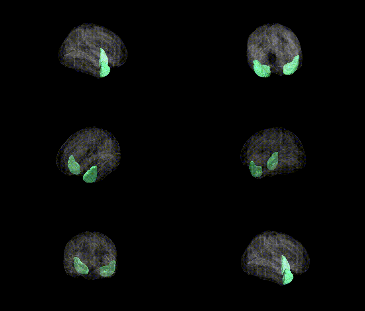
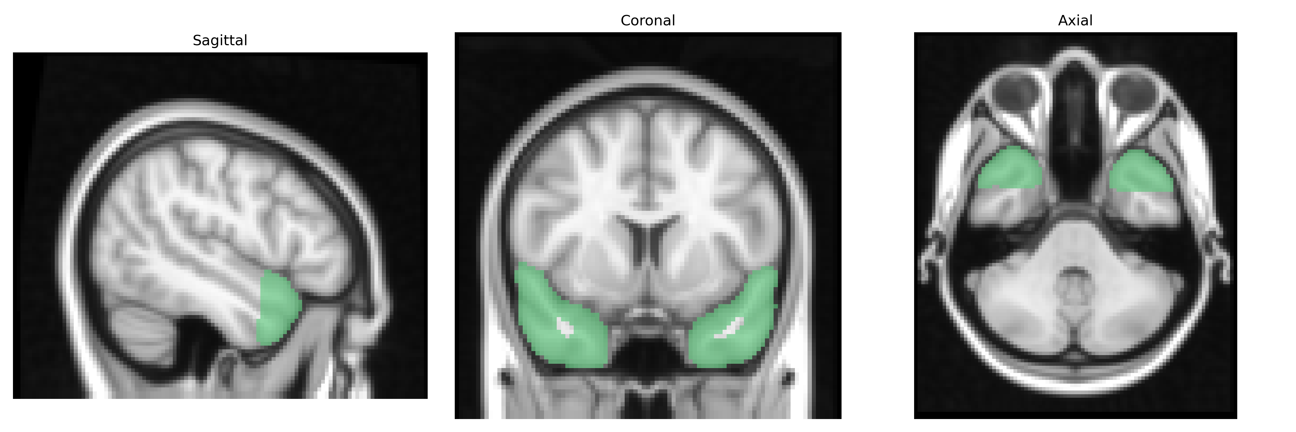
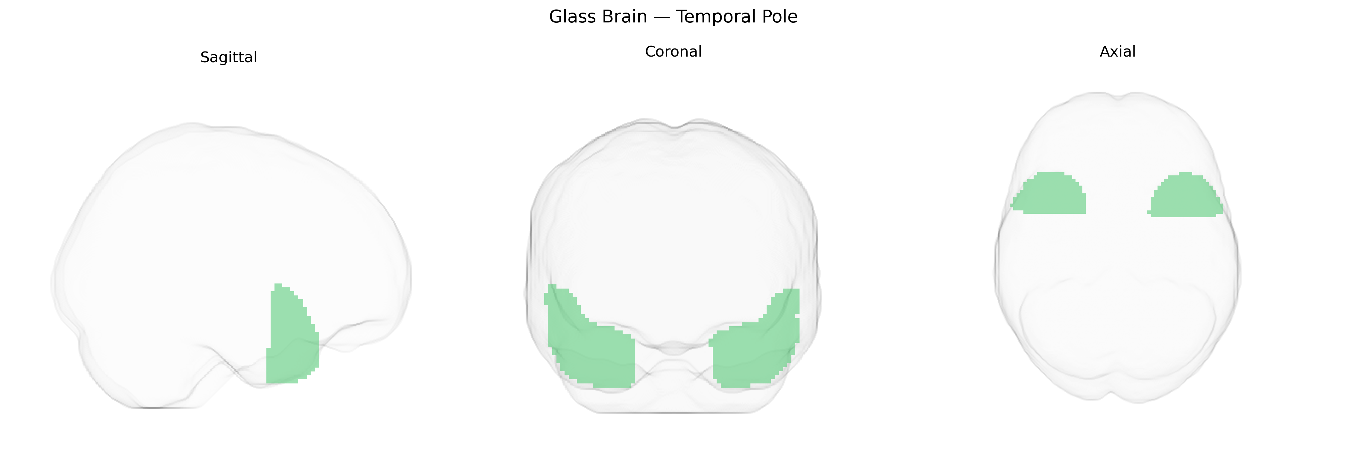
The bilateral Temporal Pole, as defined in the Harvard-Oxford cortical atlas, encompasses the most anterior portion of the temporal lobes, located rostral to the temporal neocortex and lying anterior to the temporal horn of the lateral ventricle. This region includes agranular and dysgranular cortex that is strongly interconnected with limbic, paralimbic, and higher-order association areas, including the orbitofrontal cortex, amygdala, hippocampal formation, and superior temporal regions. Functionally, the Temporal Pole is implicated in multimodal semantic processing, social and emotional cognition, autobiographical memory, and integration of complex sensory and affective information, and it is frequently involved in temporal lobe epilepsy and frontotemporal lobar degeneration. There is no direct Wikipedia article for the “Temporal Pole” as a standalone entry; a closely related structure and functional context is described under Temporal lobe.
The bilateral temporal poles, as defined in the Harvard-Oxford Cortical Atlas (maxprob thr25 2mm), have been implicated in a range of genetic and genome-wide association findings primarily through their roles in social cognition, semantic memory, and emotion processing. Structural and functional variation in this region has been linked to polygenic risk for major psychiatric disorders, including schizophrenia, bipolar disorder, and major depressive disorder, with several large-scale imaging genetics consortia (e.g., ENIGMA, UK Biobank–based GWAS) reporting associations between temporal pole thickness or volume and common risk variants in genes related to synaptic plasticity, neurodevelopment, and calcium signaling (such as CACNA1C, GRIN2A/B, and other schizophrenia/bipolar risk loci). Temporal pole atrophy shows strong genetic overlap with frontotemporal dementia, particularly variants in MAPT, GRN, and C9orf72, which are associated with degeneration of anterior temporal regions and semantic-variant primary progressive aphasia. Additional GWAS work has linked temporal pole morphology and connectivity to polygenic scores for autism spectrum disorder and social-behavioral traits, implicating genes involved in excitatory–inhibitory balance and axonal guidance, although individual locus-level findings are typically modest and highly polygenic. Overall, genetic associations with the temporal pole emerge not as region-specific single-gene effects, but as part of distributed, pleiotropic architectures influencing large-scale temporal and limbic networks implicated in emotion, language, and social cognition.
Overview generated by GPT-4o (2026).
Region ID: 8
Hemisphere: bilateral
Atlas: HarvardOxford cort maxprob thr25 2mm
Full Quality Version: Download MP4
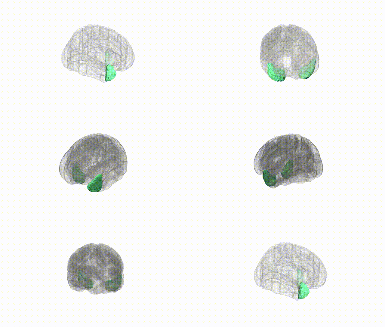
Full Quality Version: Download MP4
Wali Sidiqyar*, Gaurav Rudravaram*, Elyssa M. McMaster, Trent M. Schwartz, Adam M. Saunders, Kurt G. Schilling, Bennett A. Landman "Introducing SPINS: A Shared Public Visualization Library of Neuroanatomical Structures." Medical Imaging with Deep Learning- short paper
This resource is licensed under CC0 1.0 Universal (Public Domain).