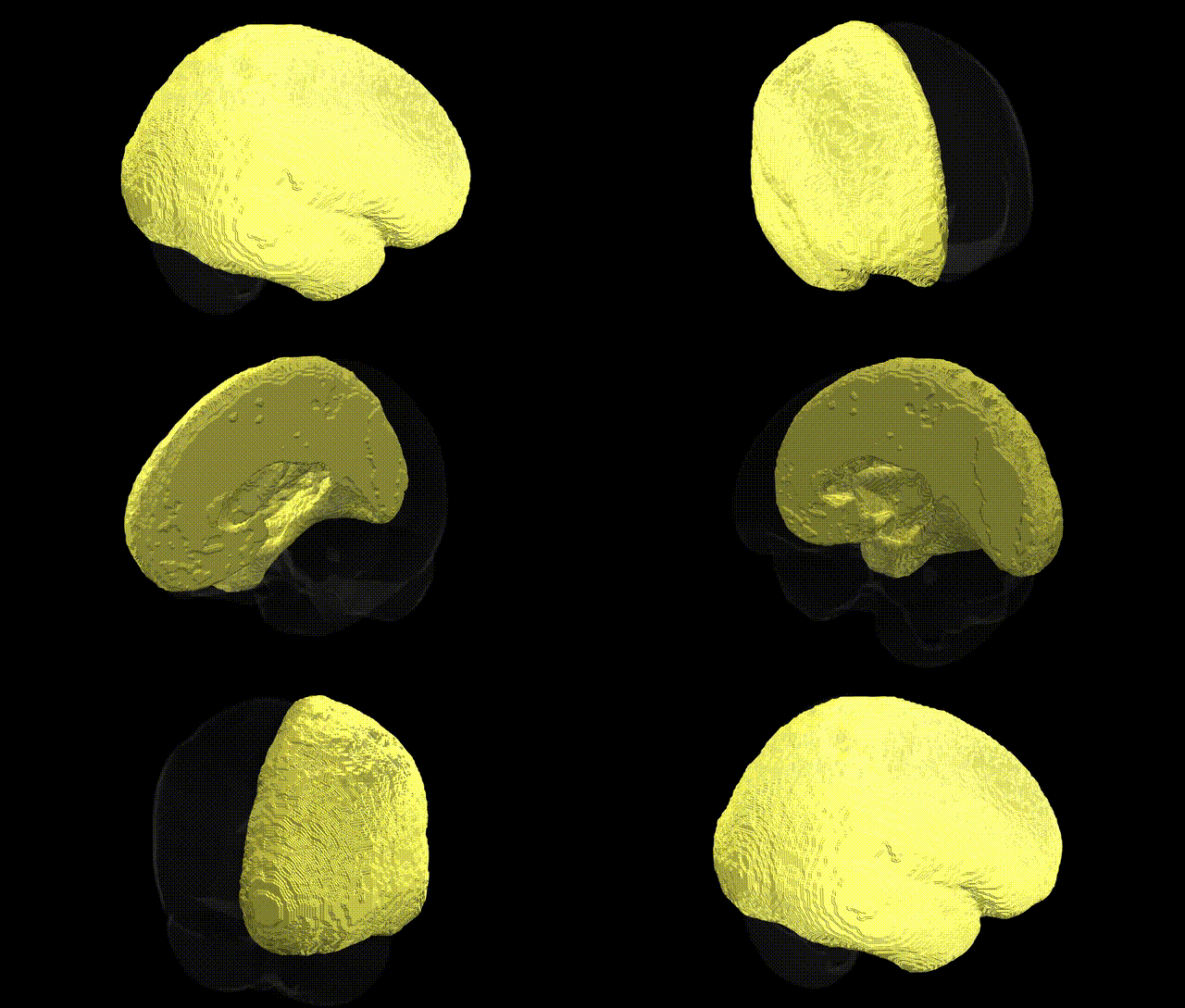
The bilateral Left Cerebral Cortex in the Harvard-Oxford subcortical maximum probability (25% threshold, 1 mm) atlas refers to the ensemble of cortical gray matter regions located within the left cerebral hemisphere, encompassing frontal, parietal, temporal, and occipital lobes as defined in volumetric atlas space. This structure includes primary and association areas involved in higher-order cognitive functions such as language, executive control, memory, sensory integration, and voluntary motor planning, although specific functional specializations vary across constituent cortical subregions. In neuroimaging and morphometric analyses, this region is typically used as a gross anatomical mask or reference compartment for parcellation, laterality assessment, and normalization of regional measurements. There is no direct link for “Left Cerebral Cortex” as a distinct article; a related structure is the Cerebral Cortex.
The bilateral left cerebral cortex as defined by the Harvard–Oxford atlas encompasses widespread association, sensory, and motor territories, and its genetic architecture is highly polygenic, with many loci showing regionally specific rather than cortex-wide effects; nevertheless, several broad findings recur across GWAS and imaging–genetics studies. Large consortia such as ENIGMA and UK Biobank have identified variants near genes involved in neurodevelopment (e.g., HMGA2, LRP6, MIR137, TBR1, and genes in Wnt and Notch signaling) that influence total and regional cortical thickness, surface area, and gyrification in left-hemisphere-dominant regions including frontal, temporal, and parietal cortex, often with modest lateralization favoring the left side for language and executive networks. Polygenic risk for schizophrenia, bipolar disorder, and major depression, as well as specific loci in genes such as CACNA1C, GRIN2A, and ZNF804A, have been associated with structural and functional alterations across left prefrontal and temporal cortex, including reduced cortical thickness and altered connectivity in default-mode and frontotemporal networks. Variants linked to neurodevelopmental traits (e.g., FOXP2, DCDC2, CNTNAP2) show associations with language-related left perisylvian cortex and reading/language disorders, while Alzheimer’s disease risk loci (e.g., APOE ε4, CLU, PICALM) have been tied to accelerated cortical thinning in left temporal and parietal association regions. Additionally, large-scale GWAS of cognition and educational attainment implicate distributed left cortical regions through polygenic scores, with genes involved in synaptic plasticity, axon guidance, and neuronal proliferation contributing to interindividual variability in cortical morphology and functional organization within the broadly defined bilateral left cerebral cortex.
Overview generated by GPT-4o (2026).
Region ID: 2
Hemisphere: bilateral
Atlas: HarvardOxford sub maxprob thr25 1mm
Full Quality Version: Download MP4
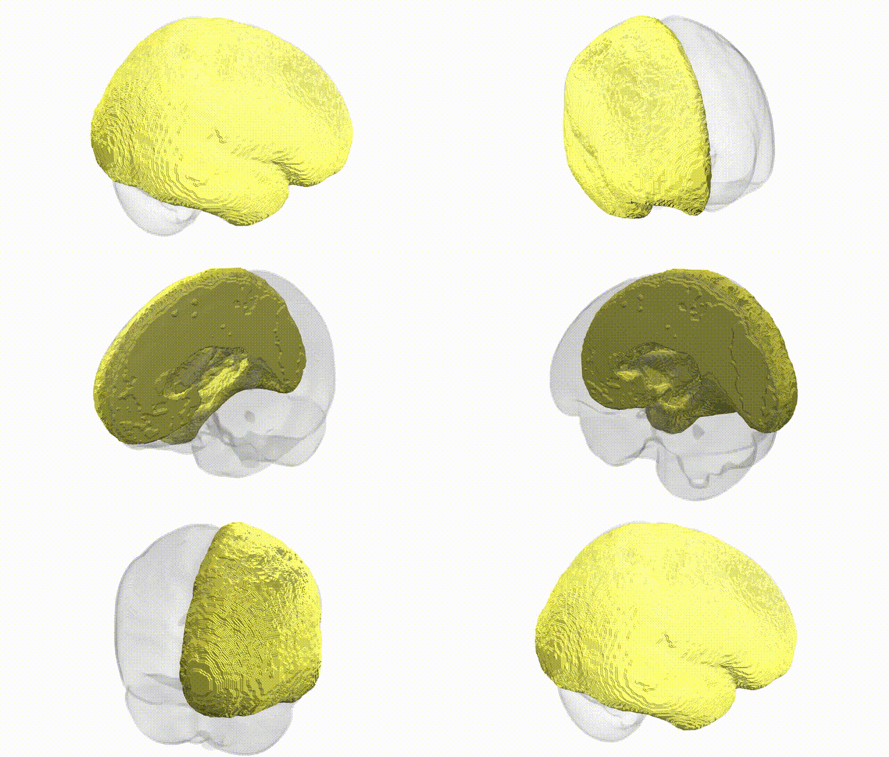
Full Quality Version: Download MP4
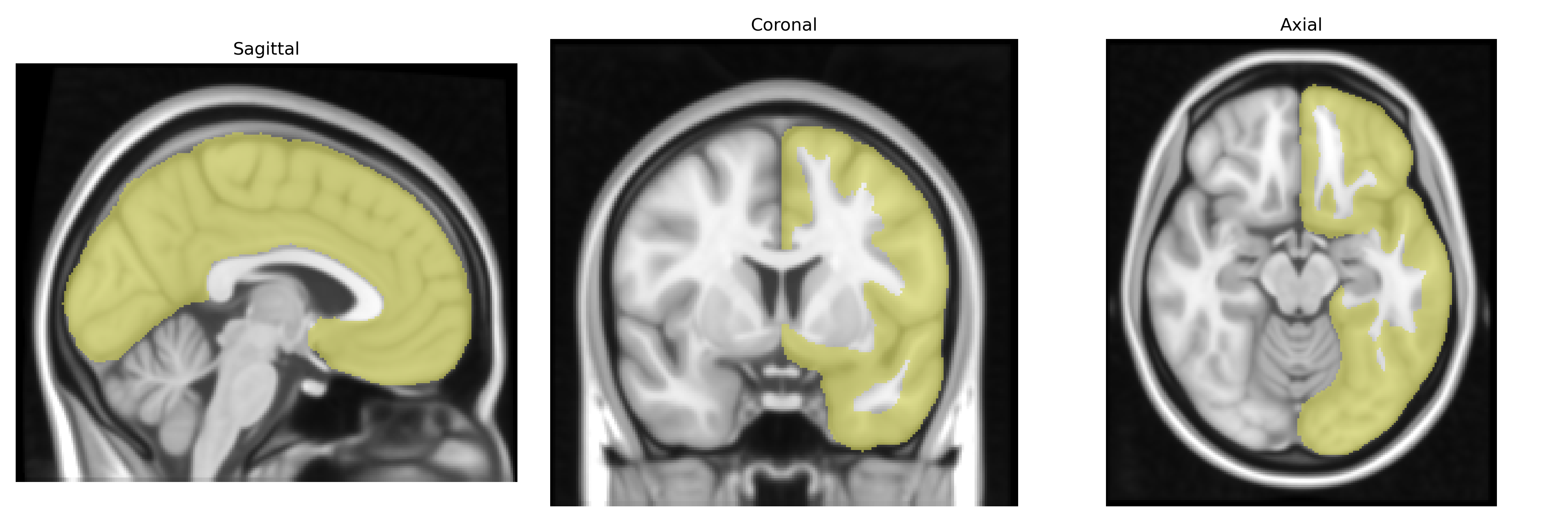
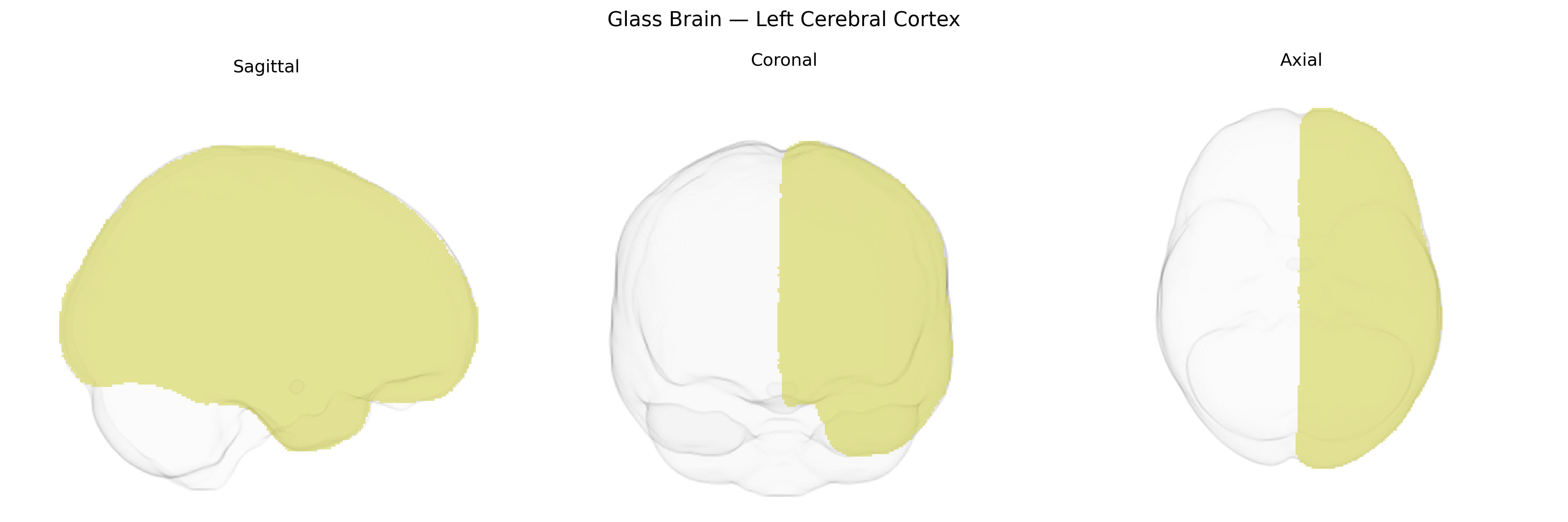
Wali Sidiqyar*, Gaurav Rudravaram*, Elyssa M. McMaster, Trent M. Schwartz, Adam M. Saunders, Kurt G. Schilling, Bennett A. Landman "Introducing SPINS: A Shared Public Visualization Library of Neuroanatomical Structures." Medical Imaging with Deep Learning- short paper
This resource is licensed under CC0 1.0 Universal (Public Domain).