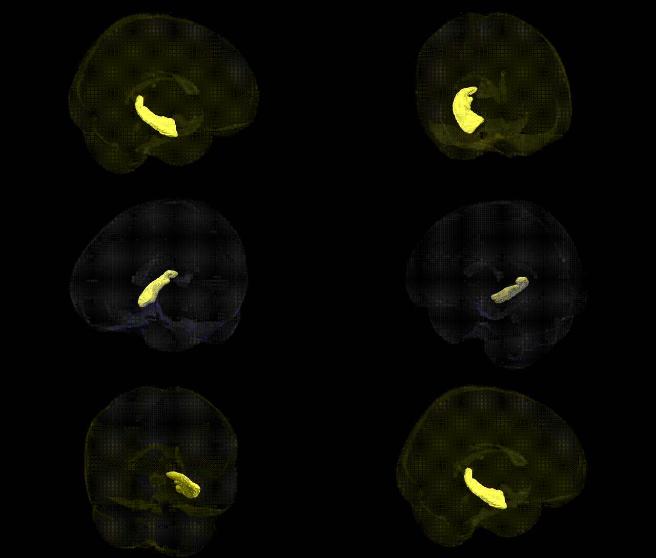
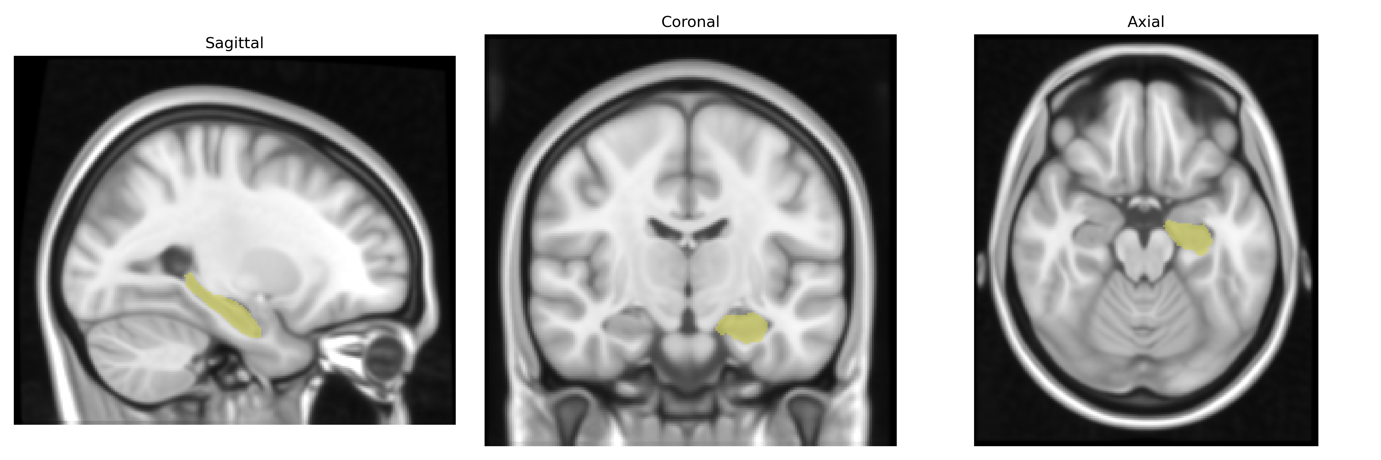
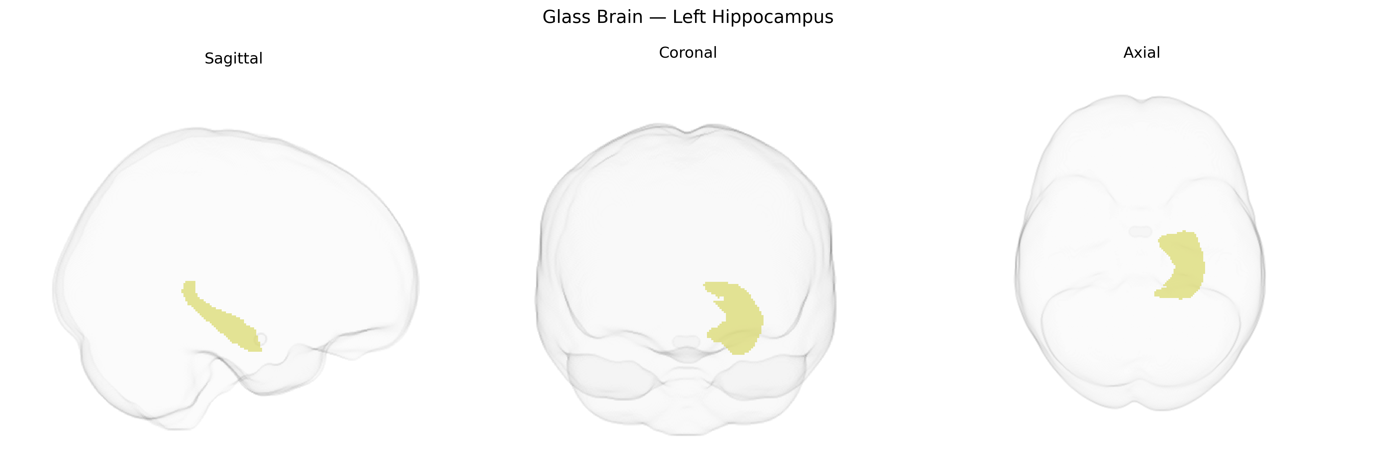
The bilateral Left Hippocampus, as defined in the Harvard–Oxford subcortical maximum probability (thr25, 1 mm) atlas, refers to the left-hemispheric portion of the hippocampal formation, a key medial temporal lobe structure involved in episodic memory encoding and retrieval, spatial navigation, and contextual learning. Cytoarchitectonically, it comprises subfields including CA1–CA4, dentate gyrus, and subiculum, and receives multimodal input via the entorhinal cortex while projecting through the fornix to diencephalic and cortical targets, notably the mammillary bodies and prefrontal regions. Functionally, the left hippocampus is often associated with verbal and narrative memory processing, and its structural integrity and activity patterns are critical in neurodegenerative conditions (e.g., Alzheimer’s disease), temporal lobe epilepsy, and affective disorders. A closely related overview of this structure is available at Hippocampus.
The bilateral left hippocampus, as defined in the Harvard-Oxford subcortical atlas, has been implicated in numerous genetic association studies, particularly large-scale GWAS of subcortical brain volumes and neuropsychiatric traits. Common variants in genes such as HMGA2, ASTN2, DPP4, VRK2, KIAA0586, and others have been associated with hippocampal volume, with several loci showing lateralized or hemisphere-specific effects, though many influence bilateral hippocampal structure broadly. GWAS meta-analyses (e.g., ENIGMA) have identified multiple SNPs associated with hippocampal volume that overlap with risk loci for Alzheimer’s disease (e.g., near APOE and BIN1), schizophrenia, major depressive disorder, and bipolar disorder, suggesting shared genetic architecture between hippocampal morphology and psychiatric/neurodegenerative disorders. Hippocampal volume–linked variants also intersect with genes involved in neurodevelopment, synaptic plasticity, and stress response, and have been tied to cognitive traits such as memory performance, general intelligence, and educational attainment. Additionally, polygenic risk scores for disorders including Alzheimer’s disease, schizophrenia, and depression have been associated with reduced hippocampal volume, particularly in the left hippocampus, supporting a role for this region as a structural mediator of genetic risk on cognition and psychopathology.
Overview generated by GPT-4o (2026).
Region ID: 9
Hemisphere: bilateral
Atlas: HarvardOxford sub maxprob thr25 1mm
Full Quality Version: Download MP4
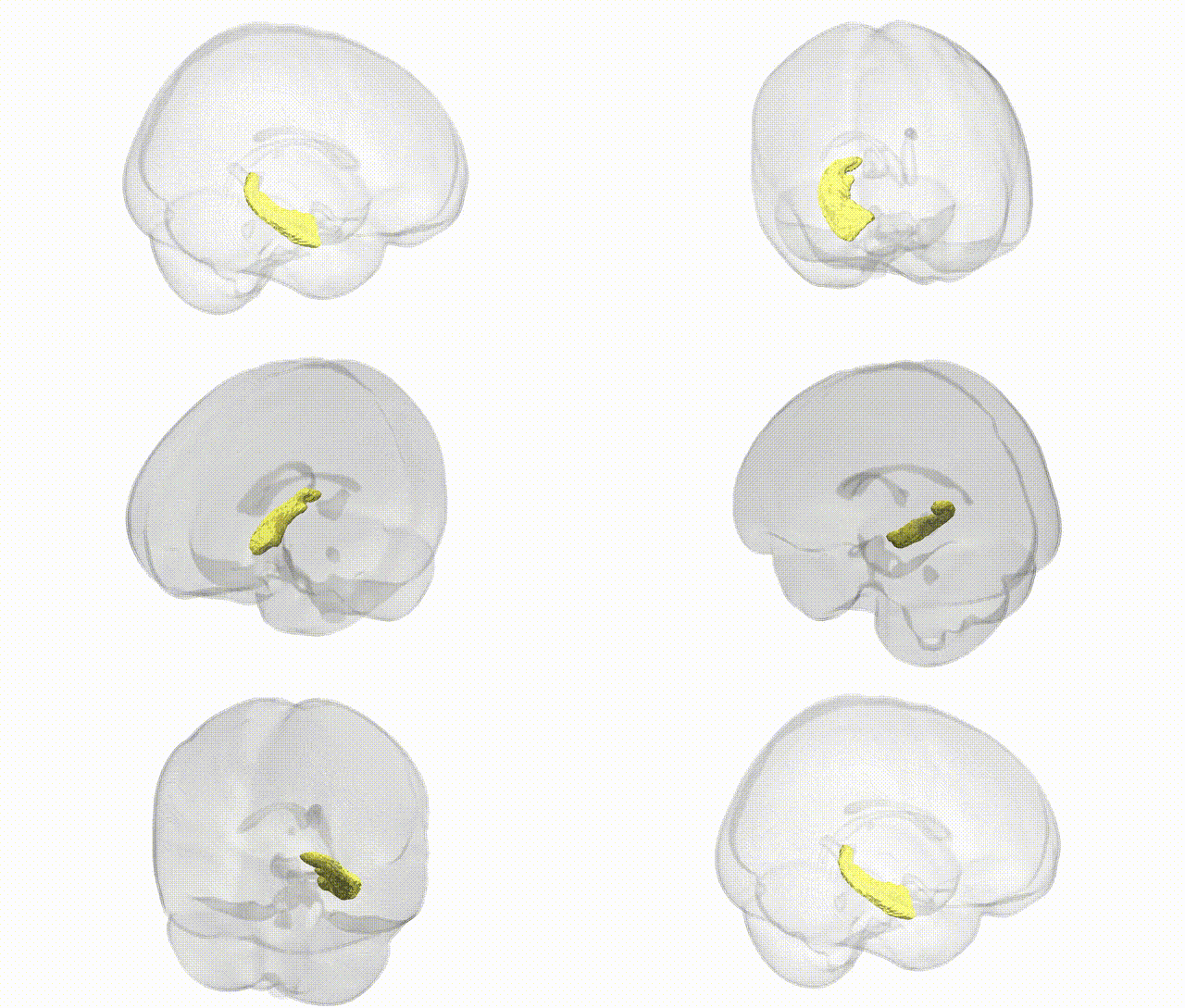
Full Quality Version: Download MP4
Wali Sidiqyar*, Gaurav Rudravaram*, Elyssa M. McMaster, Trent M. Schwartz, Adam M. Saunders, Kurt G. Schilling, Bennett A. Landman "Introducing SPINS: A Shared Public Visualization Library of Neuroanatomical Structures." Medical Imaging with Deep Learning- short paper
This resource is licensed under CC0 1.0 Universal (Public Domain).