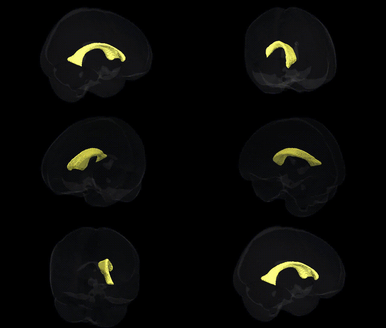
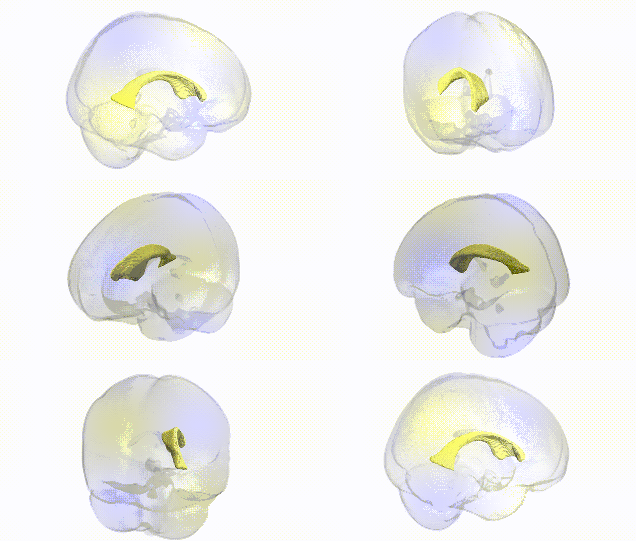
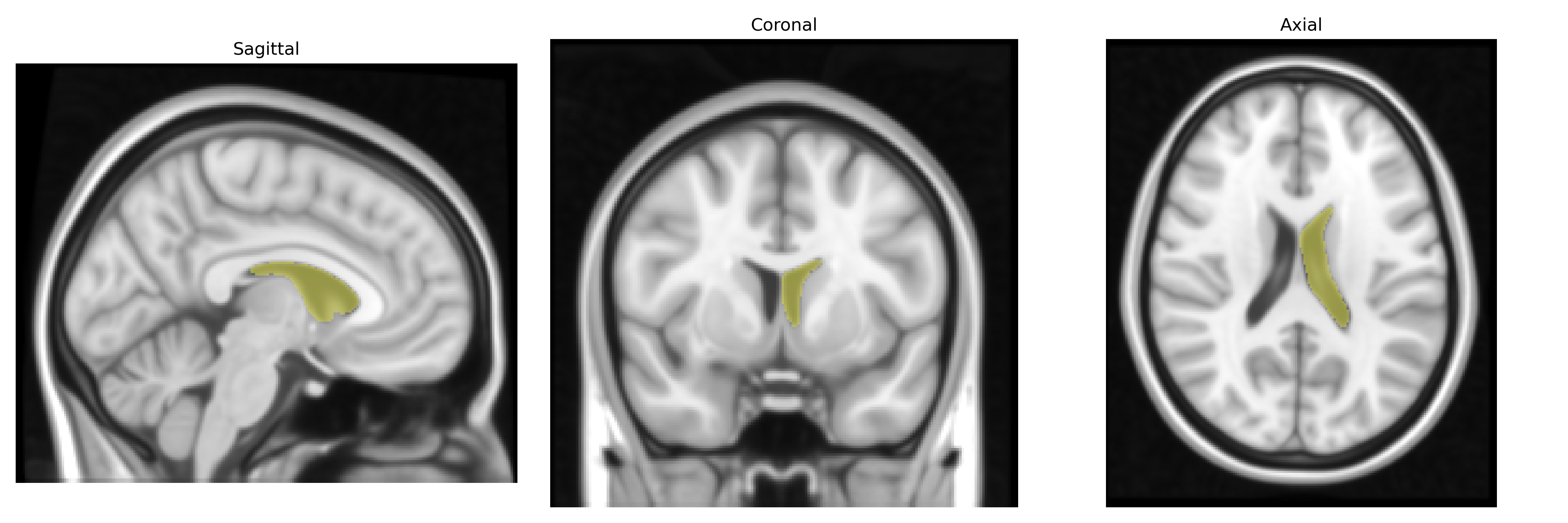
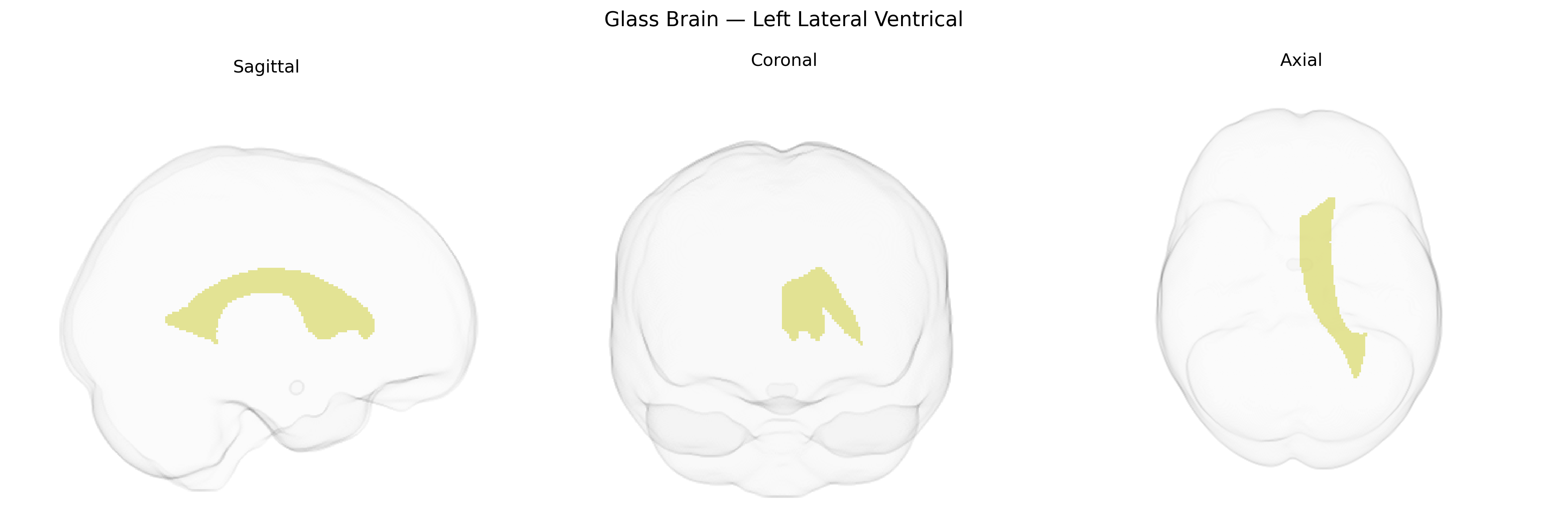
The bilateral Left Lateral Ventricle, as defined in the Harvard-Oxford subcortical atlas, corresponds to the cerebrospinal fluid-filled cavity within the left cerebral hemisphere that forms part of the paired lateral ventricles of the brain. It comprises the anterior (frontal) horn, body, posterior (occipital) horn, and inferior (temporal) horn, bordered primarily by structures of the basal ganglia, thalamus, and surrounding white matter tracts. This ventricular space is lined by ependymal cells and is continuous with the right lateral ventricle via the interventricular foramen (foramen of Monro), allowing cerebrospinal fluid to flow into the third ventricle and through the ventricular system. Functionally, the lateral ventricle participates in cerebrospinal fluid circulation, contributing to mechanical protection, buoyancy, and homeostatic regulation of the brain’s extracellular environment, and is a key anatomical landmark in neuroimaging and neurosurgical planning. Lateral ventricle
The bilateral left lateral ventricle, as defined in the Harvard‐Oxford subcortical atlas, is primarily a cerebrospinal fluid space rather than gray matter, so genetic associations identified in GWAS typically reflect ventricular volume or shape as an imaging-derived phenotype rather than a functional “region” per se. Large neuroimaging GWAS (e.g., ENIGMA, UK Biobank) have identified multiple loci influencing lateral ventricle volume, including variants near genes such as APOE, MAPT, TESC, and others involved in neurodevelopment, axonal maintenance, and neurodegeneration; enlarged lateral ventricles are consistently associated with polygenic risk for Alzheimer’s disease, schizophrenia, bipolar disorder, and major depressive disorder, as well as cognitive decline and aging-related brain atrophy. In psychiatric and neurodevelopmental GWAS integrating imaging, ventricular enlargement has been linked to higher genetic liability for schizophrenia and autism spectrum disorders, often co-localizing with risk loci affecting cortical thickness and subcortical structures rather than the ventricle itself. Studies of rare variants and monogenic disorders (e.g., hydrocephalus, certain leukodystrophies, and ciliopathy-related genes like L1CAM and CCDC88C) demonstrate that mutations in pathways controlling cerebrospinal fluid dynamics and brain morphogenesis can produce pronounced ventriculomegaly, again implicating ventricular size as a genetically influenced marker of broader neurostructural pathology. Collectively, genetic findings tie variation in lateral ventricle volume—including the left lateral ventricle—to polygenic architectures underlying neurodegeneration, psychosis, cognitive aging, and brain development, with the ventricle serving as a sensitive structural correlate rather than a primary genetic target.
Overview generated by GPT-4o (2026).
Region ID: 3
Hemisphere: bilateral
Atlas: HarvardOxford sub maxprob thr25 1mm
Full Quality Version: Download MP4
Full Quality Version: Download MP4
Wali Sidiqyar*, Gaurav Rudravaram*, Elyssa M. McMaster, Trent M. Schwartz, Adam M. Saunders, Kurt G. Schilling, Bennett A. Landman "Introducing SPINS: A Shared Public Visualization Library of Neuroanatomical Structures." Medical Imaging with Deep Learning- short paper
This resource is licensed under CC0 1.0 Universal (Public Domain).