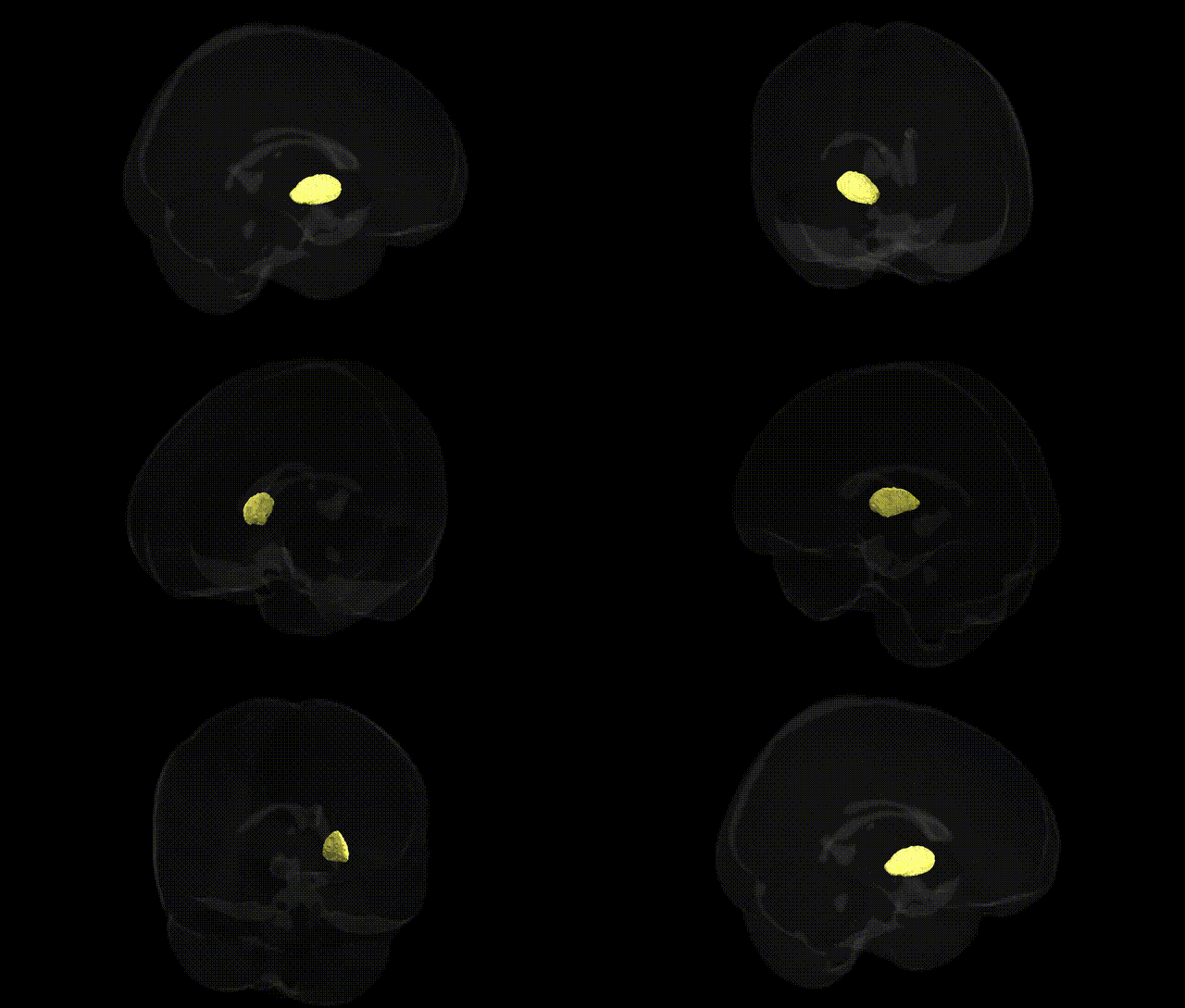
The bilateral left pallidum is the left-sided component of the globus pallidus, a core structure of the basal ganglia located deep within the cerebral hemispheres, medial to the putamen and lateral to the internal capsule. It is subdivided into an external (globus pallidus externa) and internal (globus pallidus interna) segment, which receive major input from the striatum and project to thalamic and brainstem nuclei. Through its position in direct and indirect basal ganglia pathways, the pallidum plays a central role in the regulation of voluntary movement, muscle tone, and motor learning, as well as in aspects of motivation and reward processing. Its dysfunction is implicated in movement disorders such as Parkinson’s disease and dystonia. There is no direct Wikipedia article specifically for “Left Pallidum”; a related and encompassing structure is the Globus pallidus.
The bilateral left pallidum, a core component of the basal ganglia circuitry implicated in motor control, reward processing, and habit learning, has been linked through genetic studies to multiple neuropsychiatric and behavioral phenotypes, although most GWAS interrogate pallidal volume or function rather than the specific Harvard-Oxford atlas label. Genome-wide association studies of subcortical brain volumes (e.g., ENIGMA consortium) have identified variants near genes such as KTN1, DLG2, and TMEM106B, among others, associated with pallidum size, suggesting polygenic influences on its structure. Pallidal abnormalities, often genetically mediated, are repeatedly observed in disorders like Parkinson’s disease (linked to LRRK2, SNCA, GBA), dystonia (TOR1A, GNAL), Huntington’s disease (HTT expansion), and Wilson’s disease (ATP7B), where these risk genes alter basal ganglia circuitry that prominently involves the pallidum. GWAS of schizophrenia, bipolar disorder, and major depressive disorder (implicating loci near CACNA1C, DRD2, GRIN2A, and several synaptic and neurodevelopmental genes) have been associated with pallidal volume and functional changes in imaging-genetics studies, which report shared polygenic architectures between psychiatric risk and pallidal morphology. Additionally, genetic influences on traits such as cognitive performance, impulsivity, and substance use (e.g., variants in DRD2, OPRM1, and dopamine signaling pathways) have been functionally linked to pallidal activity and connectivity, reinforcing the view that the left pallidum’s structural and functional variation arises from distributed, pleiotropic genetic effects rather than single locus determinants.
Overview generated by GPT-4o (2026).
Region ID: 7
Hemisphere: bilateral
Atlas: HarvardOxford sub maxprob thr25 1mm
Full Quality Version: Download MP4
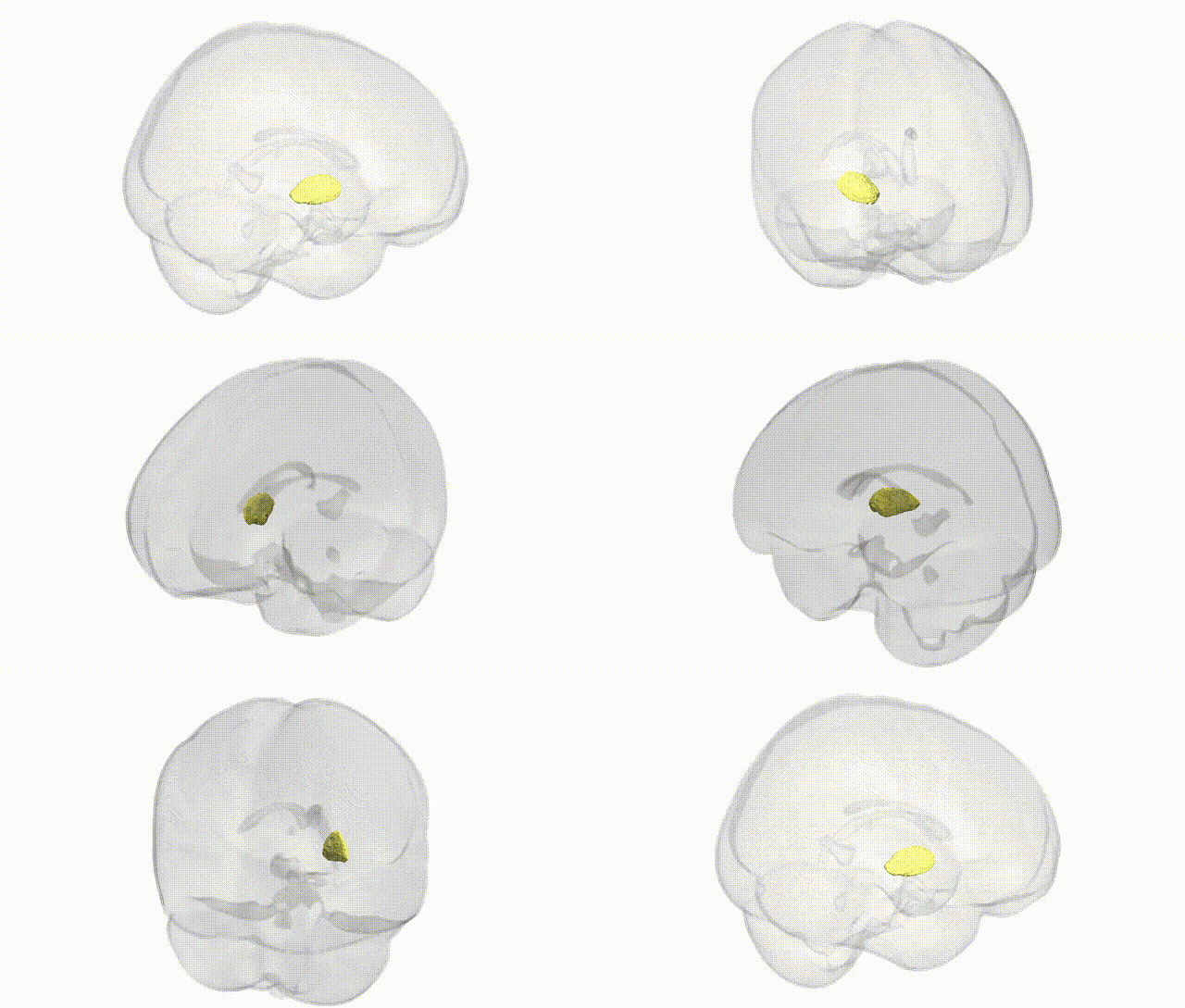
Full Quality Version: Download MP4
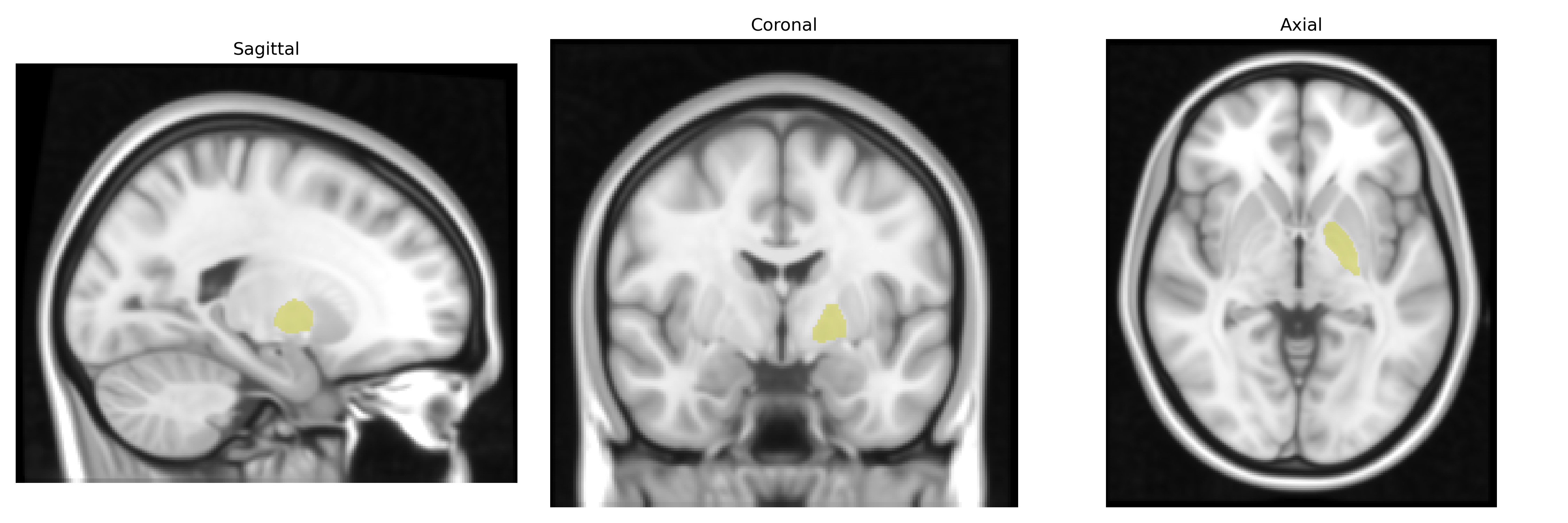
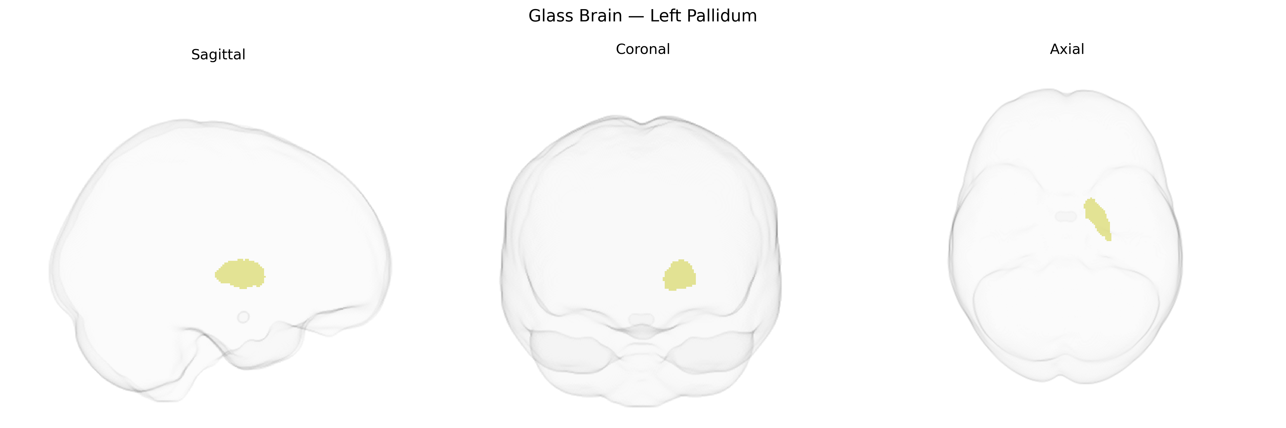
Wali Sidiqyar*, Gaurav Rudravaram*, Elyssa M. McMaster, Trent M. Schwartz, Adam M. Saunders, Kurt G. Schilling, Bennett A. Landman "Introducing SPINS: A Shared Public Visualization Library of Neuroanatomical Structures." Medical Imaging with Deep Learning- short paper
This resource is licensed under CC0 1.0 Universal (Public Domain).