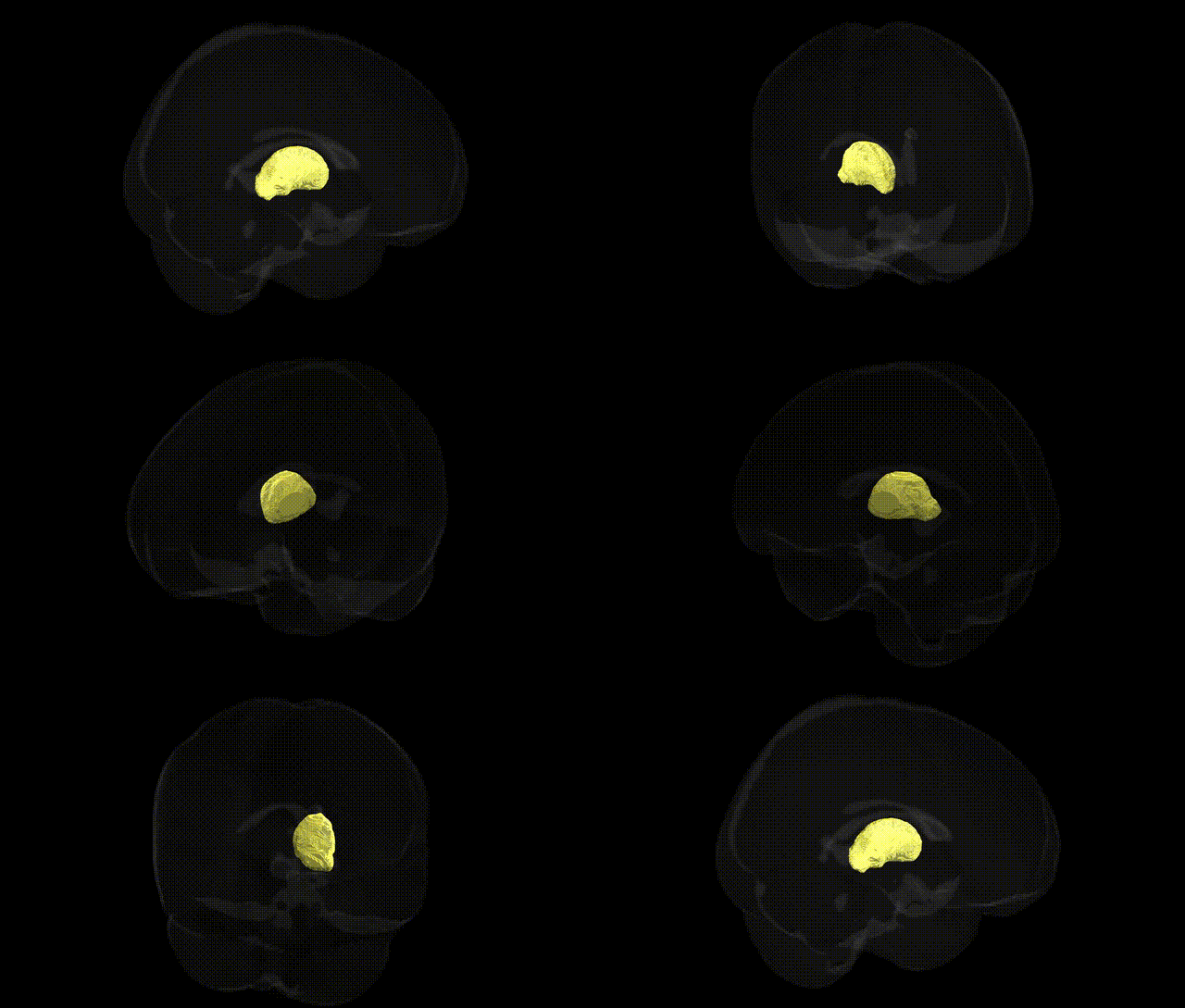
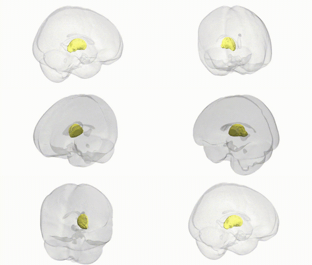
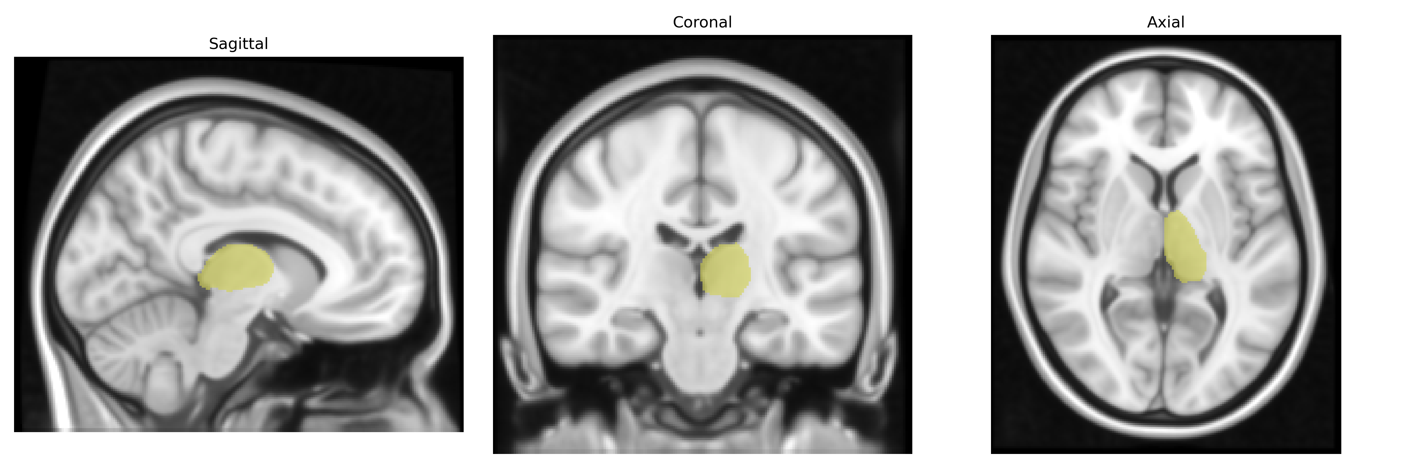
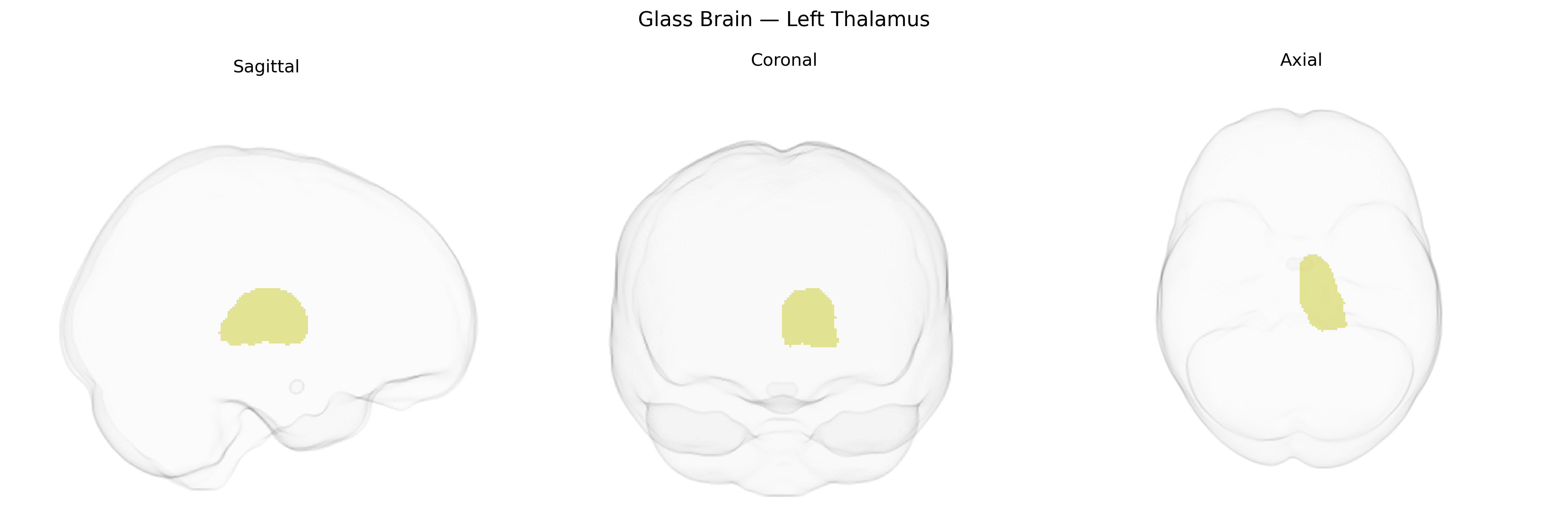
The bilateral Left Thalamus, as defined in the Harvard-Oxford subcortical atlas, comprises the left-sided portion of the thalamic complex, a paired deep grey matter structure located medial to the internal capsule and superior to the brainstem within the diencephalon. It is composed of multiple nuclei (e.g., relay, association, and intralaminar nuclei) that process and integrate ascending sensory information (excluding most olfactory input) and project to widespread cortical regions, contributing critically to sensory perception, motor control pathways, arousal, attention, and aspects of cognition and consciousness. The thalamus also participates in cortico-striato-thalamo-cortical loops implicated in motor planning, executive functions, and limbic processing, and is a key node in large-scale neural networks modulated by neuromodulatory systems. Thalamus
The bilateral left thalamus, as delineated in the Harvard-Oxford subcortical atlas, has been implicated in multiple genetic and GWAS findings linking its volume, microstructure, and function to diverse traits and disorders. Large-scale imaging–genetics consortia (e.g., ENIGMA, UK Biobank) have identified common variants in genes related to neurodevelopment, axon guidance, and synaptic signaling—including loci near or within MAPT, SLC39A8, CNTNAP2, and calcium-channel or glutamatergic pathway genes—that influence thalamic volume and connectivity. Polygenic risk scores for schizophrenia, bipolar disorder, and major depressive disorder show associations with altered thalamic volume or thalamo-cortical connectivity, while specific risk loci for schizophrenia (e.g., in the MHC region, CACNA1C-related networks) and autism spectrum disorder (genes involved in synaptic function and neuronal migration) have been linked to structural and functional thalamic differences. GWAS of cognitive performance, intelligence, and educational attainment implicate variants that also relate to thalamic size and white-matter integrity, supporting a role for thalamic genetic architecture in general cognitive function. In addition, genetic risk for neurodegenerative diseases such as Parkinson’s disease and Alzheimer’s disease, involving genes like LRRK2, GBA, and APOE, has been associated with thalamic atrophy or altered thalamic connectivity, and variants influencing vascular health and blood pressure contribute to subcortical, including thalamic, small-vessel damage. Collectively, these findings indicate that the left thalamus is a key subcortical hub whose morphology and connectivity are shaped by polygenic influences that overlap across psychiatric, cognitive, and neurodegenerative phenotypes.
Overview generated by GPT-4o (2026).
Region ID: 4
Hemisphere: bilateral
Atlas: HarvardOxford sub maxprob thr25 1mm
Full Quality Version: Download MP4
Full Quality Version: Download MP4
Wali Sidiqyar*, Gaurav Rudravaram*, Elyssa M. McMaster, Trent M. Schwartz, Adam M. Saunders, Kurt G. Schilling, Bennett A. Landman "Introducing SPINS: A Shared Public Visualization Library of Neuroanatomical Structures." Medical Imaging with Deep Learning- short paper
This resource is licensed under CC0 1.0 Universal (Public Domain).