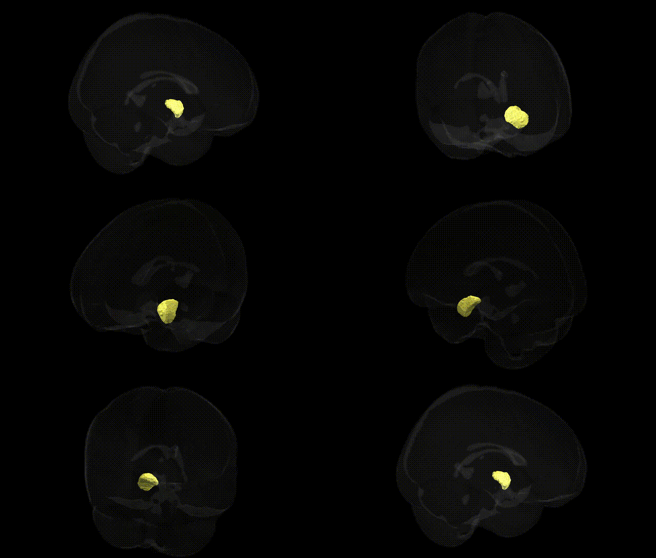
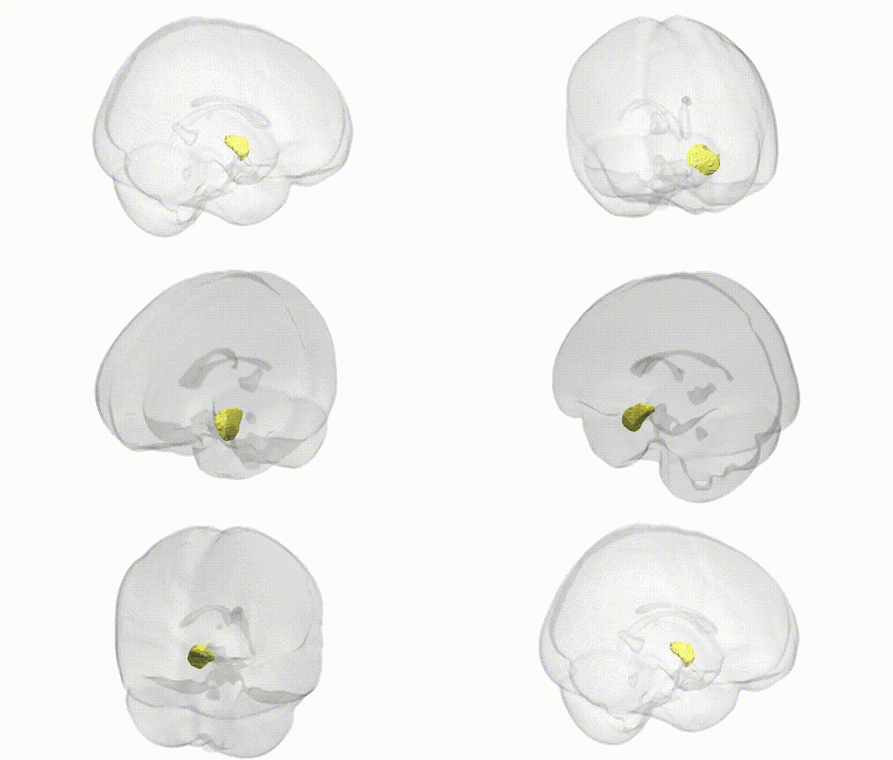
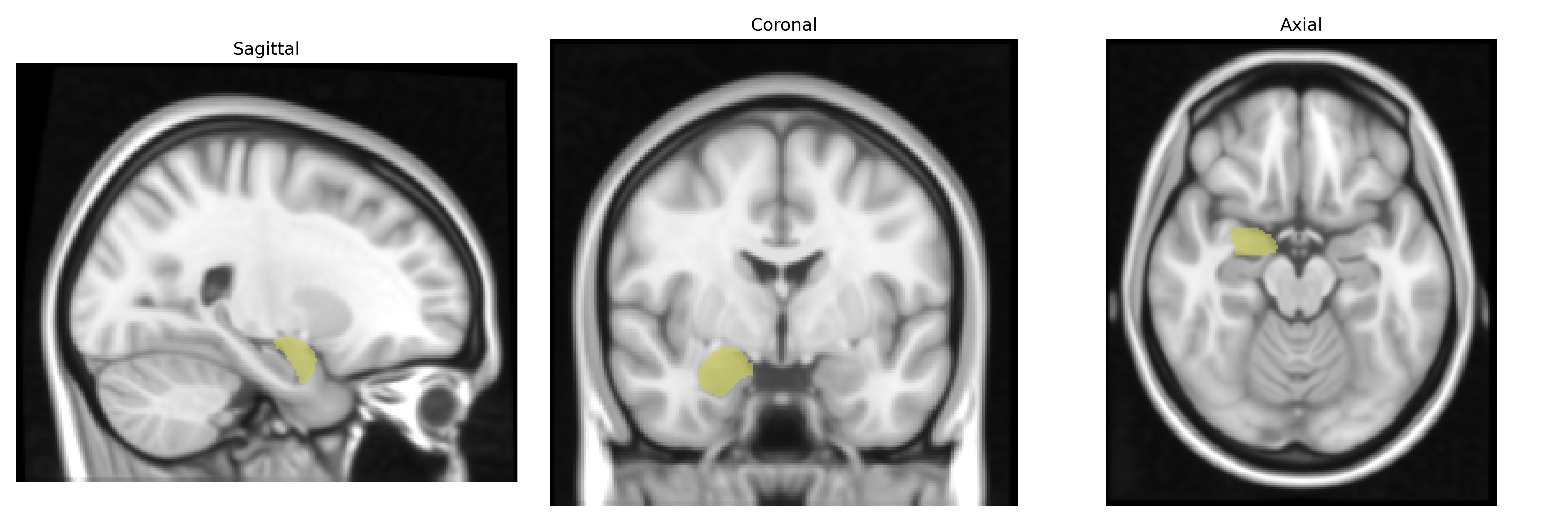
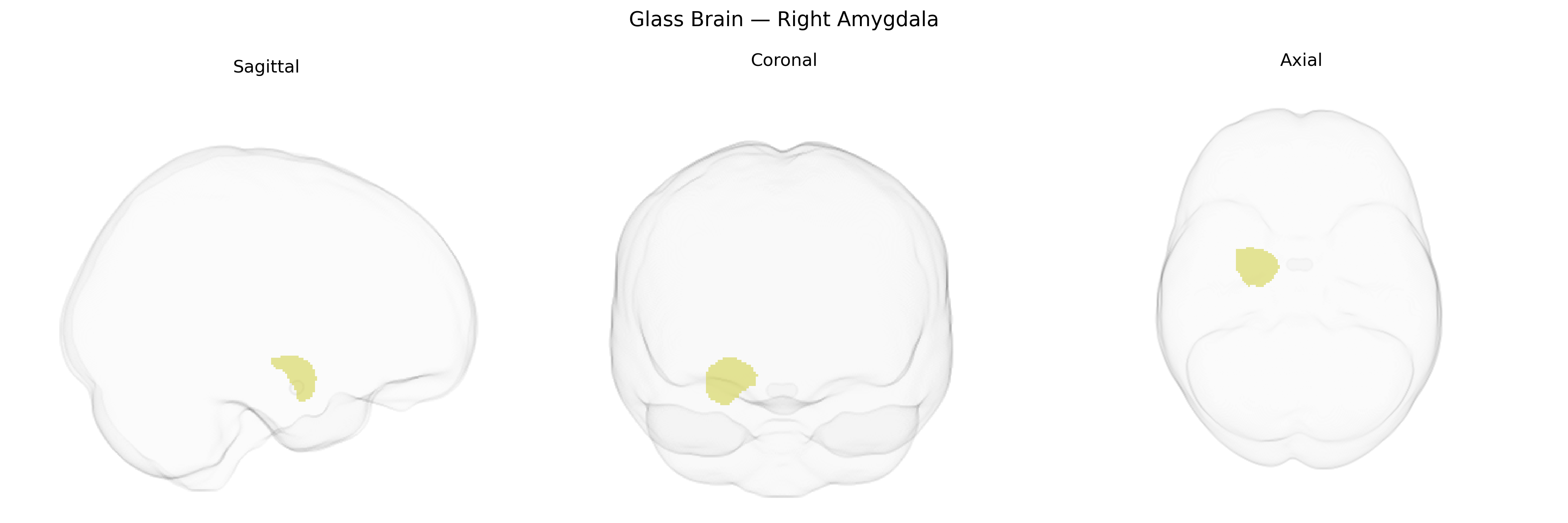
The bilateral right amygdala, as defined in the Harvard-Oxford Subcortical Atlas, is a paired, almond-shaped cluster of nuclei located in the medial temporal lobe, forming part of the limbic system and lying anterior to the hippocampus. It is primarily involved in emotional processing, particularly fear and threat detection, as well as assigning affective value to sensory stimuli and modulating autonomic and endocrine responses via extensive connections with the hypothalamus, prefrontal cortex, and brainstem. The right amygdala, in particular, has been associated with rapid, unconscious processing of emotional stimuli and may show functional asymmetries relative to the left amygdala in tasks involving vigilance, arousal, and facial emotion recognition. Structurally, it comprises several subnuclei (basolateral, centromedial, and cortical groups) that integrate multimodal sensory input and influence memory consolidation in concert with the hippocampal formation. Amygdala
Genetic associations with the bilateral right amygdala, as defined in the Harvard-Oxford subcortical atlas, largely arise from GWAS of amygdala volume and function and overlap with loci implicated in psychiatric and neurodevelopmental traits. Variants in genes involved in synaptic plasticity and neurodevelopment—including SLC6A4 (serotonin transporter), BDNF (notably Val66Met), APOE (especially ε4), COMT, and FKBP5—have been linked to right amygdala structure or reactivity, often in the context of anxiety, depression, stress-related disorders, and emotional processing. Large-scale imaging genetics studies (e.g., ENIGMA and UK Biobank–based GWAS) have identified multiple common SNPs associated with amygdala volume, with signals clustering near genes related to neuronal migration, axon guidance, and immune signaling, and these loci sometimes show pleiotropic associations with major depressive disorder, bipolar disorder, schizophrenia, PTSD, and autism spectrum traits. Functional GWAS and polygenic score analyses indicate that higher genetic liability for anxiety, neuroticism, and depression tends to be associated with altered right amygdala activation to emotional stimuli and, in some cases, reduced or asymmetrically altered amygdala volume. Additional associations include links between right amygdala morphology and genetic risk for Alzheimer’s disease (APOE region and other AD loci) and with traits such as aggression, impulsivity, and social behavior, although many findings are modest in effect size and partially overlapping with those reported for the left amygdala or total amygdala volume rather than strictly lateralized to the right side.
Overview generated by GPT-4o (2026).
Region ID: 20
Hemisphere: bilateral
Atlas: HarvardOxford sub maxprob thr25 1mm
Full Quality Version: Download MP4
Full Quality Version: Download MP4
Wali Sidiqyar*, Gaurav Rudravaram*, Elyssa M. McMaster, Trent M. Schwartz, Adam M. Saunders, Kurt G. Schilling, Bennett A. Landman "Introducing SPINS: A Shared Public Visualization Library of Neuroanatomical Structures." Medical Imaging with Deep Learning- short paper
This resource is licensed under CC0 1.0 Universal (Public Domain).