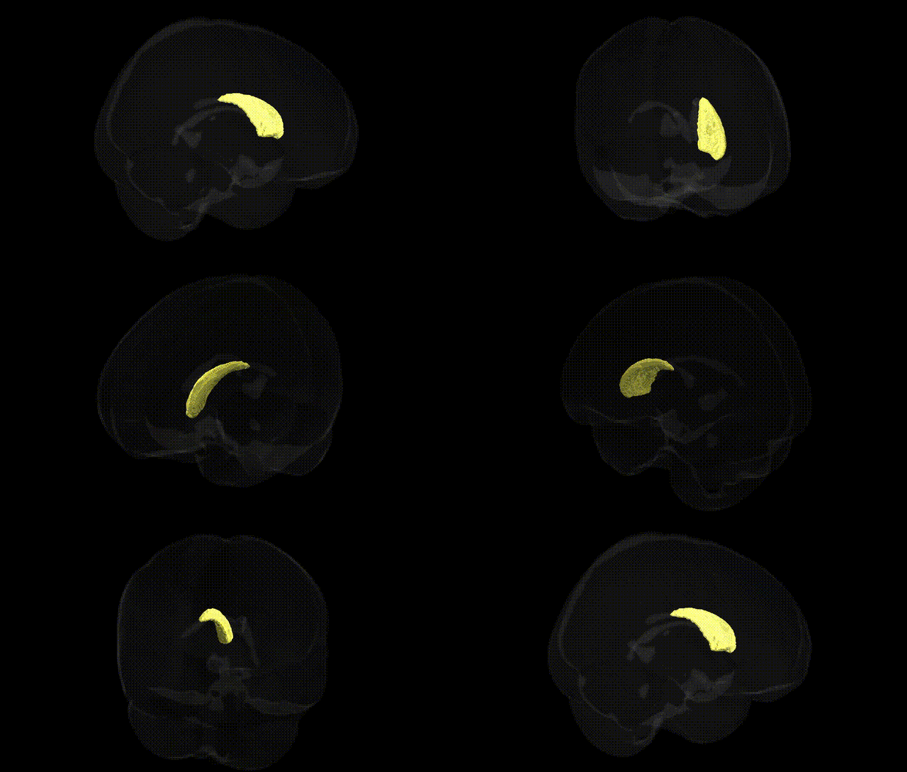
The bilateral Right Caudate, as defined in the Harvard-Oxford subcortical maxprob thr25 1mm atlas, corresponds to the right-sided component of the caudate nucleus, a C-shaped gray matter structure in the dorsal striatum of the basal ganglia. It lies adjacent to the lateral ventricle and is cytoarchitectonically continuous with the left caudate across the midline, together contributing to cortico-basal ganglia-thalamo-cortical loops involved in motor control, action selection, procedural learning, and aspects of cognition such as working memory and goal-directed behavior. The right caudate receives dense glutamatergic input from frontal and association cortices and dopaminergic input from the midbrain, integrating these signals to modulate thalamic output and influence cortical activity. Functionally, lateralized properties have been reported, with the right caudate often implicated in spatial attention and certain executive processes, though its core role is shared with the left caudate as part of the striatal network. Caudate nucleus
The bilateral right caudate, as defined in the Harvard–Oxford subcortical atlas, has been implicated in multiple genetic studies linking variation in dopamine, glutamate, and synaptic plasticity pathways to structural and functional differences in this region, with downstream associations to neuropsychiatric and cognitive phenotypes. GWAS and imaging-genetics analyses have identified common variants near or within genes such as DRD2, COMT, BDNF, CACNA1C, and GRIN2B that correlate with caudate volume, connectivity, or activity, often in the context of broader basal ganglia circuits. Polygenic risk for schizophrenia, bipolar disorder, major depressive disorder, ADHD, and obsessive–compulsive disorder shows associations with caudate morphology and function, and specific risk loci for these disorders have been linked to altered striatal signaling and corticostriatal loop integrity. Large-scale consortia (e.g., ENIGMA) report that genetic variants influencing intracranial volume, height, and general neurodevelopment also affect caudate size, highlighting shared genetic architecture between brain structure and body growth. In movement disorders, risk alleles for Huntington’s disease (HTT CAG expansion) and Parkinson’s disease (e.g., in LRRK2 and GBA) involve degeneration or dysfunction within striatal structures including the caudate, while addiction-related GWAS implicate variants in dopaminergic and opioid genes that modulate reward-related activation of the caudate. Autism spectrum conditions, Tourette syndrome, and learning-related traits have likewise been linked through GWAS and candidate-gene studies to genes affecting synaptic development and neurotransmission in striatal circuits, with the right caudate frequently emerging as a key locus of genetically influenced variability in habit formation, procedural learning, and goal-directed behavior.
Overview generated by GPT-4o (2026).
Region ID: 16
Hemisphere: bilateral
Atlas: HarvardOxford sub maxprob thr25 1mm
Full Quality Version: Download MP4
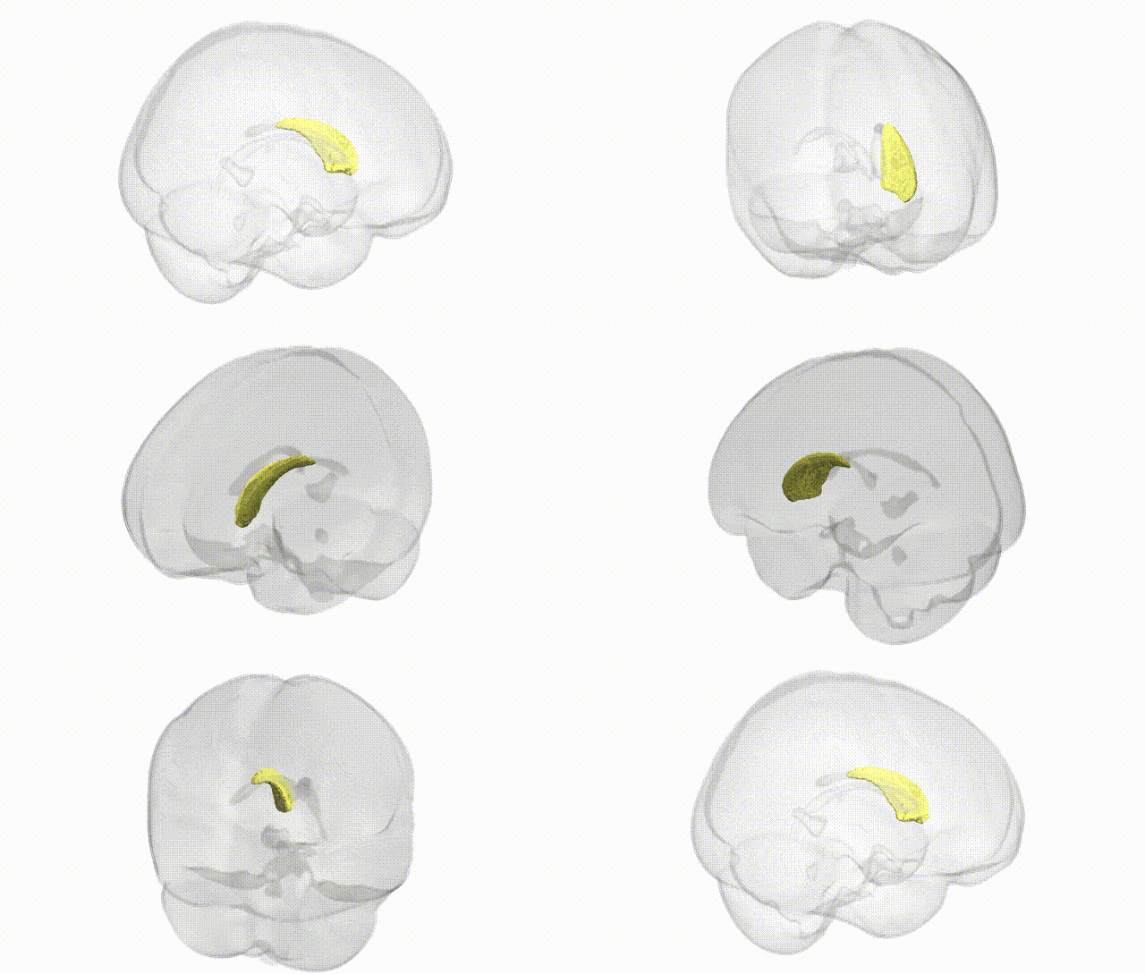
Full Quality Version: Download MP4
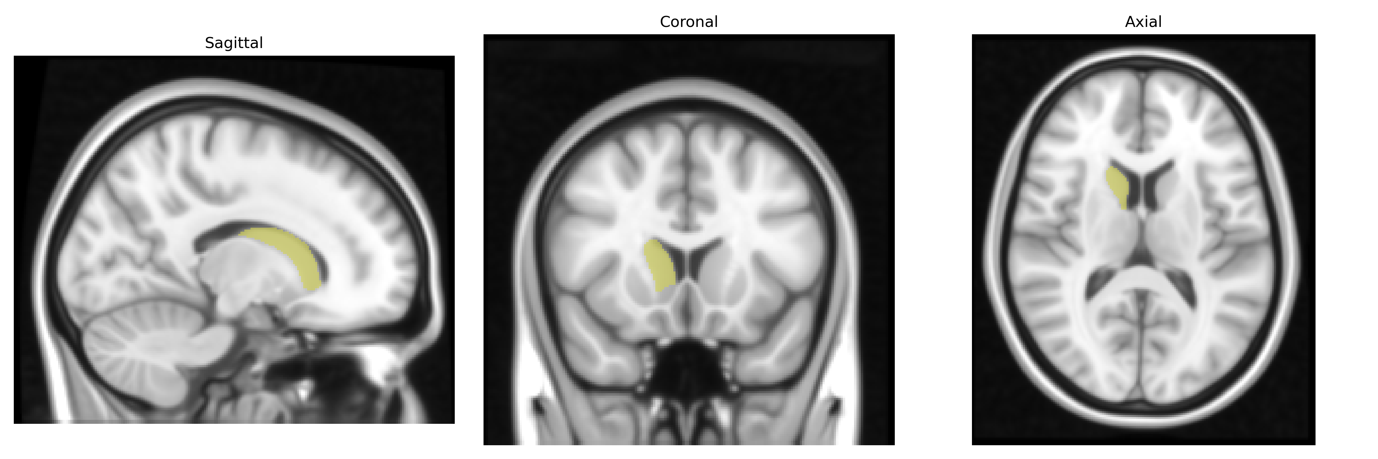
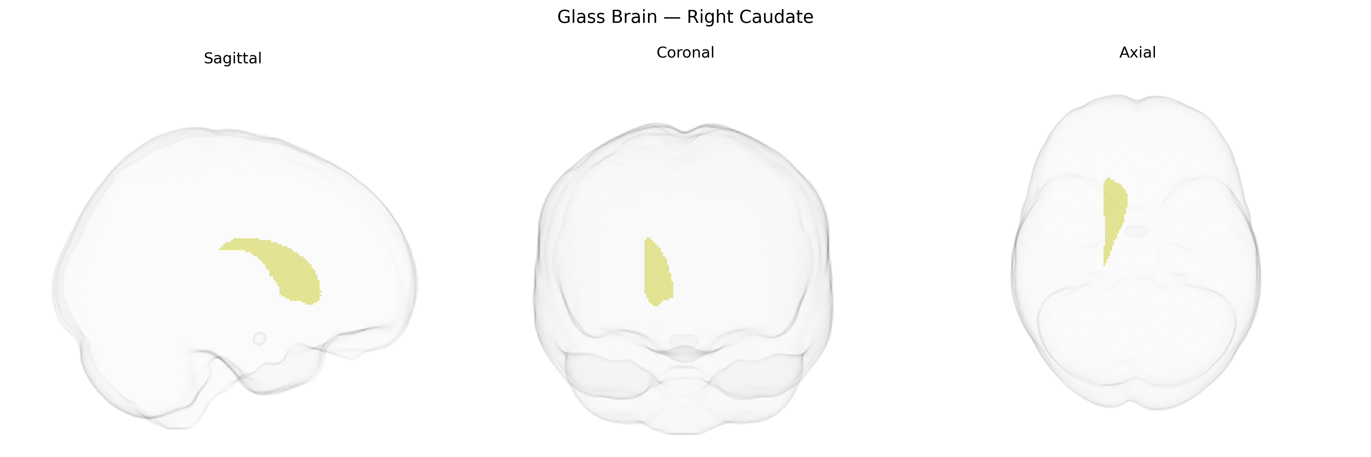
Wali Sidiqyar*, Gaurav Rudravaram*, Elyssa M. McMaster, Trent M. Schwartz, Adam M. Saunders, Kurt G. Schilling, Bennett A. Landman "Introducing SPINS: A Shared Public Visualization Library of Neuroanatomical Structures." Medical Imaging with Deep Learning- short paper
This resource is licensed under CC0 1.0 Universal (Public Domain).