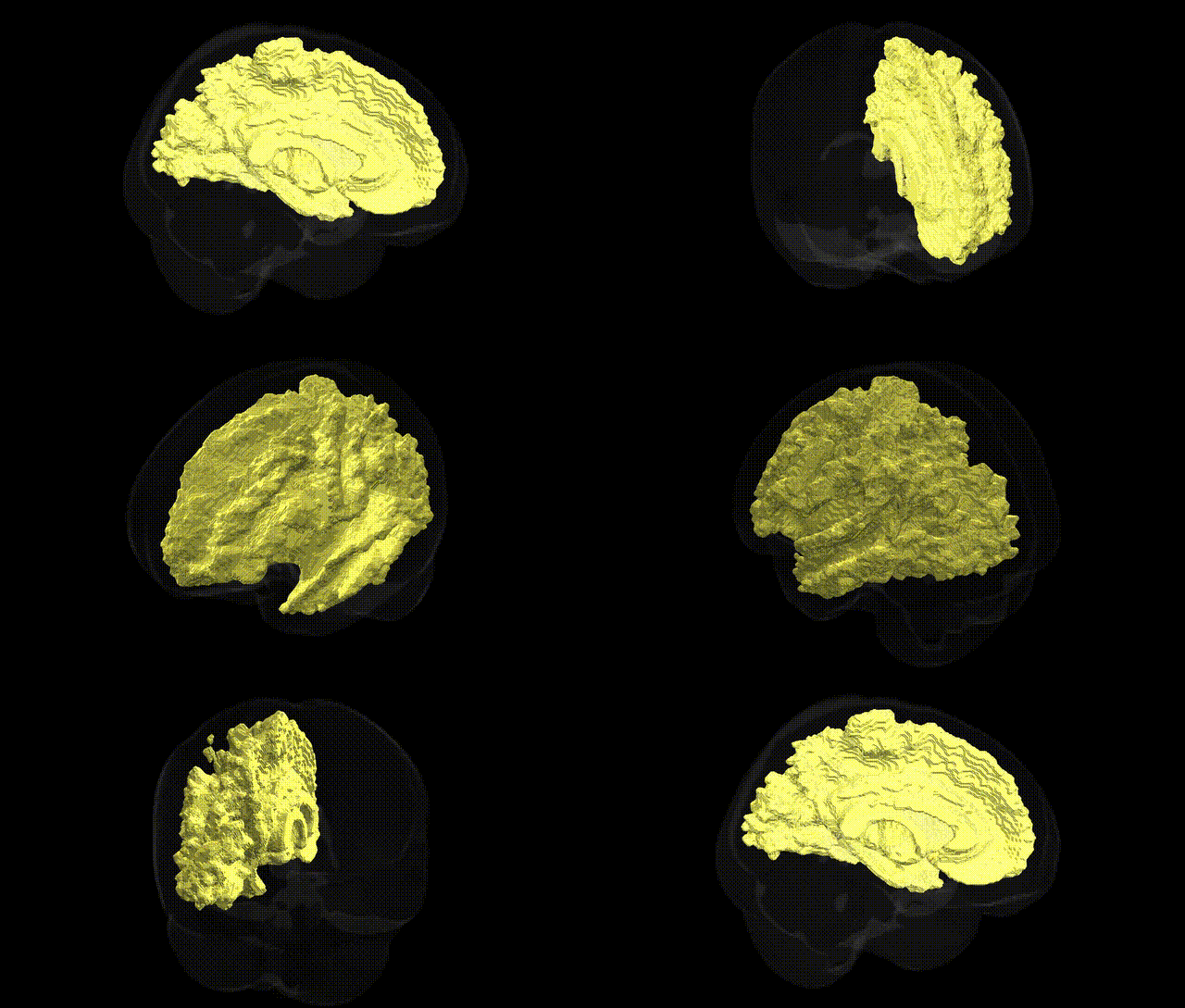
The bilateral Right Cerebral White Matter, as defined in the Harvard-Oxford subcortical atlas, comprises the myelinated fiber tracts underlying the right cerebral cortex, including association, commissural, and projection pathways that facilitate rapid signal transmission between cortical areas, across hemispheres, and between cortex and subcortical structures. This region includes major bundles such as portions of the corpus callosum, internal capsule, and long association fibers, supporting integrative functions across sensory, motor, and cognitive domains by enabling efficient communication within and beyond the right hemisphere. Cerebral white matter integrity is critical for normal brain connectivity, and its disruption is implicated in a wide range of neurological and psychiatric conditions, including demyelinating diseases, stroke, and diffuse axonal injury. There is no direct link specifically for “Right Cerebral White Matter”; a related structure is White matter.
The bilateral right cerebral white matter, as defined in the Harvard–Oxford subcortical atlas, does not have many GWAS that target this exact labeled region, but numerous imaging-genetics studies have identified genetic variants associated with global and regional white matter microstructure, which would encompass it. Common variants in genes such as APOE (especially ε4), CNTNAP2, NRG1, and DISC1 have been linked to white matter integrity and connectivity metrics derived from diffusion tensor imaging, including fractional anisotropy and mean diffusivity across widespread tracts, while loci near DCC, BDNF, and myelin-related genes (e.g., MAG, MBP) have also been implicated in white matter phenotypes. Large GWAS from consortia such as ENIGMA and UK Biobank have associated numerous SNPs with white matter volume and diffusion metrics, revealing polygenic influences and substantial genetic overlap with psychiatric and neurological disorders, including schizophrenia, bipolar disorder, major depression, autism spectrum disorder, Alzheimer’s disease, and multiple sclerosis, all of which show alterations in cerebral white matter on MRI. These findings collectively suggest that the right cerebral white matter is influenced by a distributed set of genetic variants affecting axonal development, myelination, and neuroinflammation, and that its structural characteristics share genetic risk with diverse cognitive, behavioral, and neurodegenerative traits, even though specific GWAS rarely isolate this atlas-defined region in isolation.
Overview generated by GPT-4o (2026).
Region ID: 12
Hemisphere: bilateral
Atlas: HarvardOxford sub maxprob thr25 1mm
Full Quality Version: Download MP4
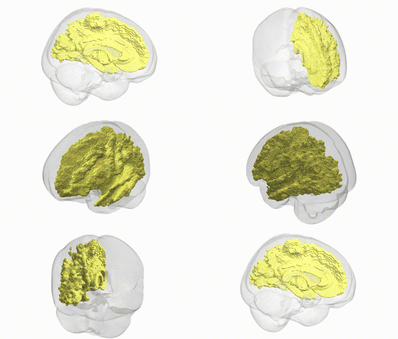
Full Quality Version: Download MP4
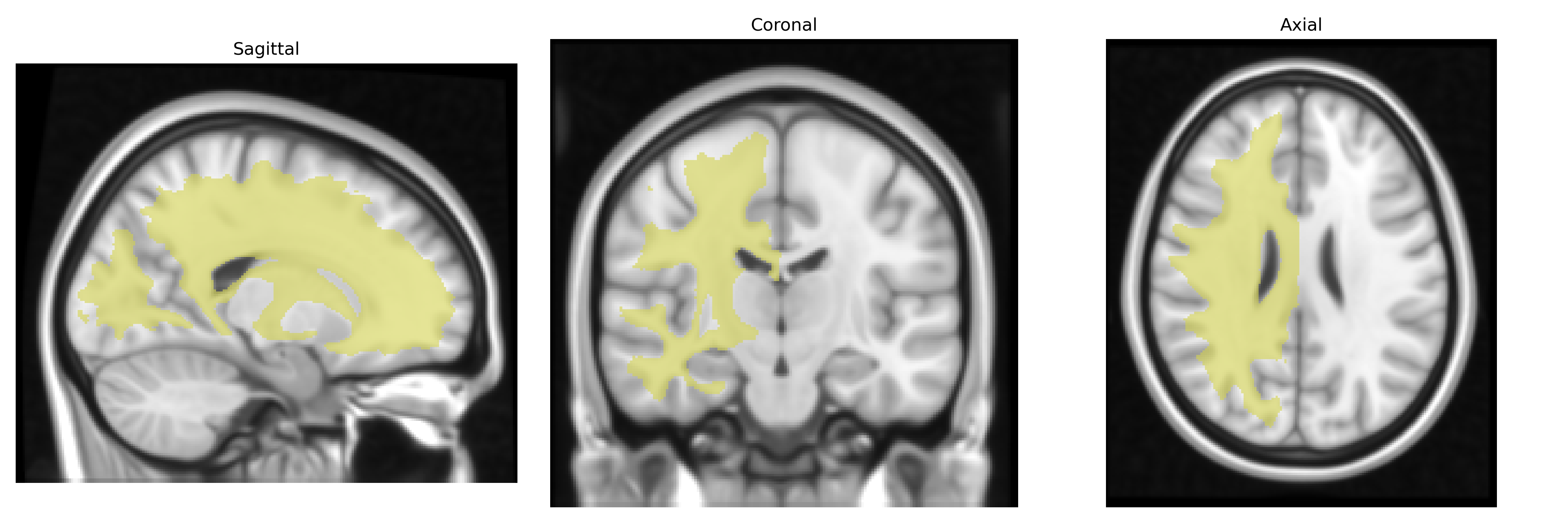
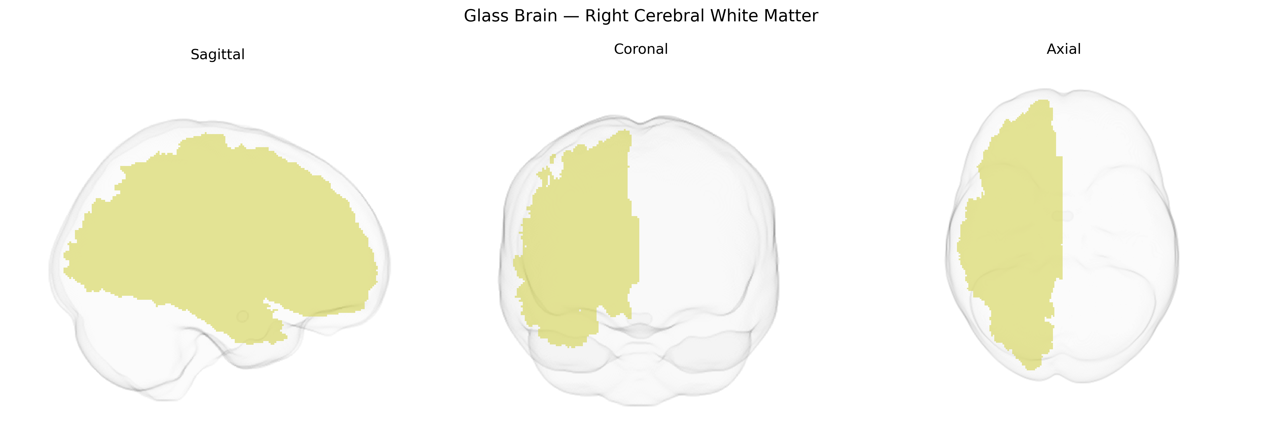
Wali Sidiqyar*, Gaurav Rudravaram*, Elyssa M. McMaster, Trent M. Schwartz, Adam M. Saunders, Kurt G. Schilling, Bennett A. Landman "Introducing SPINS: A Shared Public Visualization Library of Neuroanatomical Structures." Medical Imaging with Deep Learning- short paper
This resource is licensed under CC0 1.0 Universal (Public Domain).