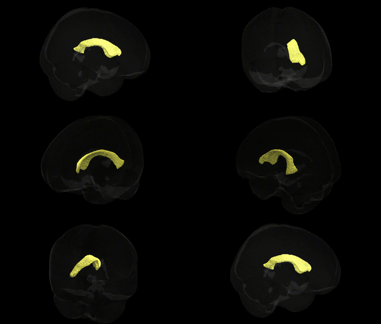
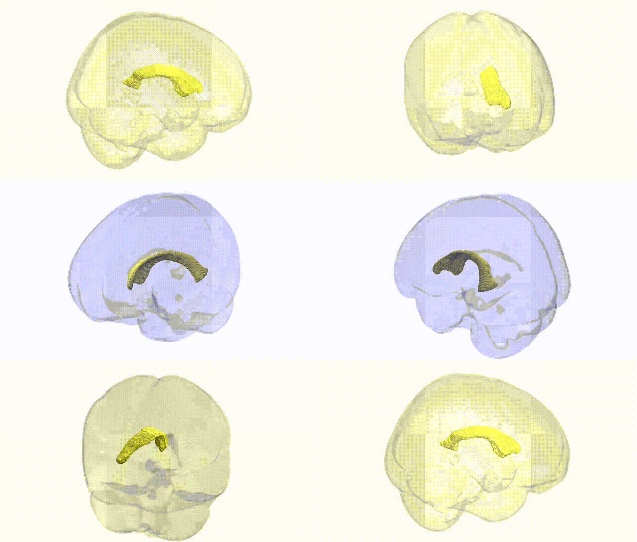
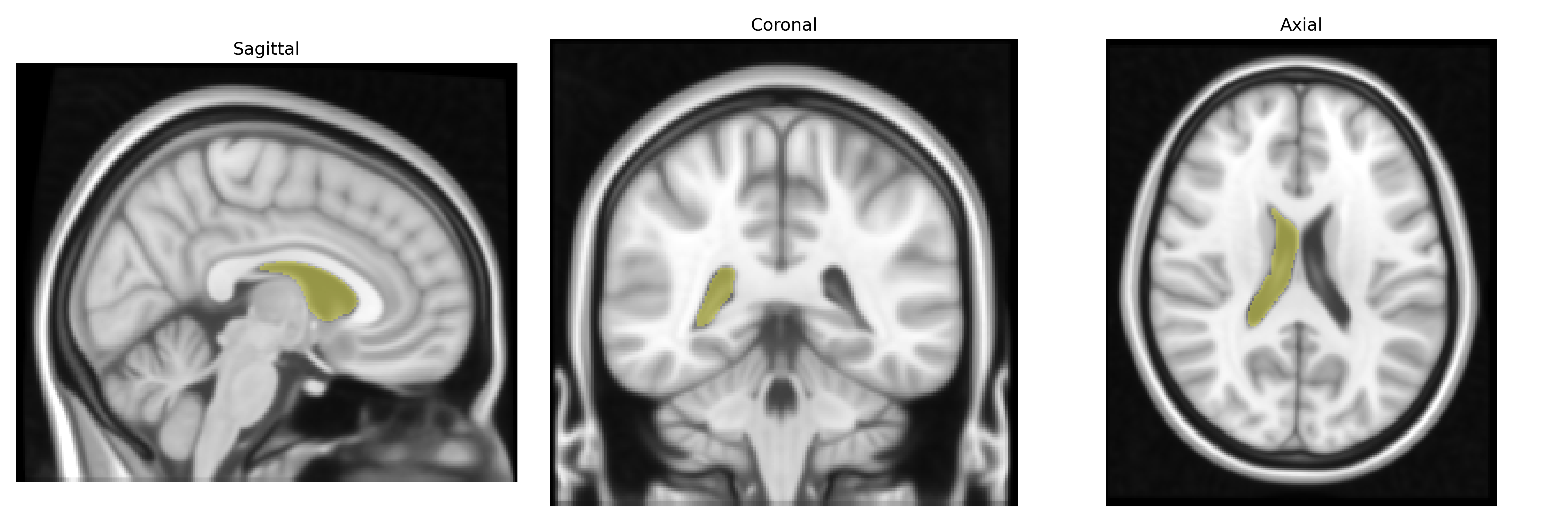
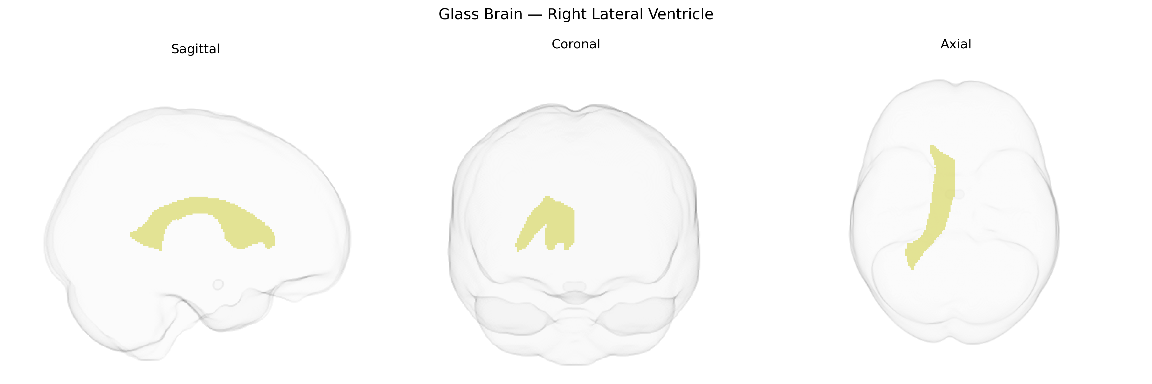
The bilateral Right Lateral Ventricle, as defined in the Harvard-Oxford subcortical atlas (maxprob thr25, 1 mm), corresponds to the right-sided component of the lateral ventricular system, a CSF-filled cavity within the telencephalon that participates in cerebrospinal fluid production, circulation, and homeostasis. It comprises the frontal (anterior) horn, body, occipital (posterior) horn, and temporal (inferior) horn, each following adjacent cortical and subcortical structures such as the caudate nucleus, corpus callosum, thalamus, and hippocampus. The ependymal lining of this ventricle and its association with the choroid plexus contribute to neurochemical regulation, metabolic waste clearance, and mechanical cushioning of brain tissue. Although a dedicated article for “Right Lateral Ventricle” does not exist, the structure is encompassed within the broader Lateral ventricle entry.
The bilateral right lateral ventricle, as defined in the Harvard–Oxford subcortical atlas, serves primarily as a cerebrospinal fluid space rather than gray matter, so genetic associations identified in GWAS typically relate to ventricular volume or shape rather than the ventricle per se. Large neuroimaging GWAS, including ENIGMA and UK Biobank studies, have shown that lateral ventricular volume is a highly heritable structural trait, with common variants in or near genes such as APOE, MAPT, and genes influencing neurodegeneration, brain aging, and cerebrospinal fluid dynamics associated with individual differences in ventricular size. Increased lateral ventricle volume has robust genetic correlations with Alzheimer’s disease, other dementias, and global brain atrophy, and has also been linked to schizophrenia and bipolar disorder through polygenic risk and imaging–genetics analyses, where risk alleles for these disorders associate with enlarged ventricles and altered surrounding white matter. In addition, GWAS of intracranial volume and subcortical structures have identified loci (e.g., in HMGA2 and other growth-related genes) that indirectly affect lateral ventricle dimensions via effects on total brain size and cortical or subcortical tissue loss, making ventricular volume a genetically influenced biomarker of neurodevelopmental and neurodegenerative processes rather than a direct target tissue itself.
Overview generated by GPT-4o (2026).
Region ID: 14
Hemisphere: bilateral
Atlas: HarvardOxford sub maxprob thr25 1mm
Full Quality Version: Download MP4
Full Quality Version: Download MP4
Wali Sidiqyar*, Gaurav Rudravaram*, Elyssa M. McMaster, Trent M. Schwartz, Adam M. Saunders, Kurt G. Schilling, Bennett A. Landman "Introducing SPINS: A Shared Public Visualization Library of Neuroanatomical Structures." Medical Imaging with Deep Learning- short paper
This resource is licensed under CC0 1.0 Universal (Public Domain).