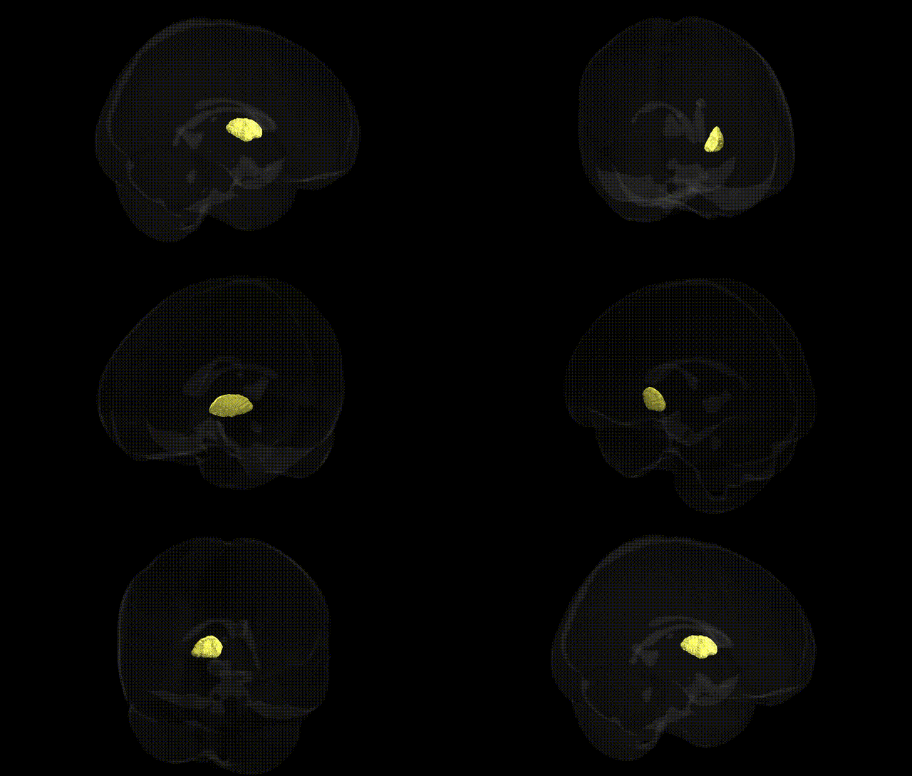
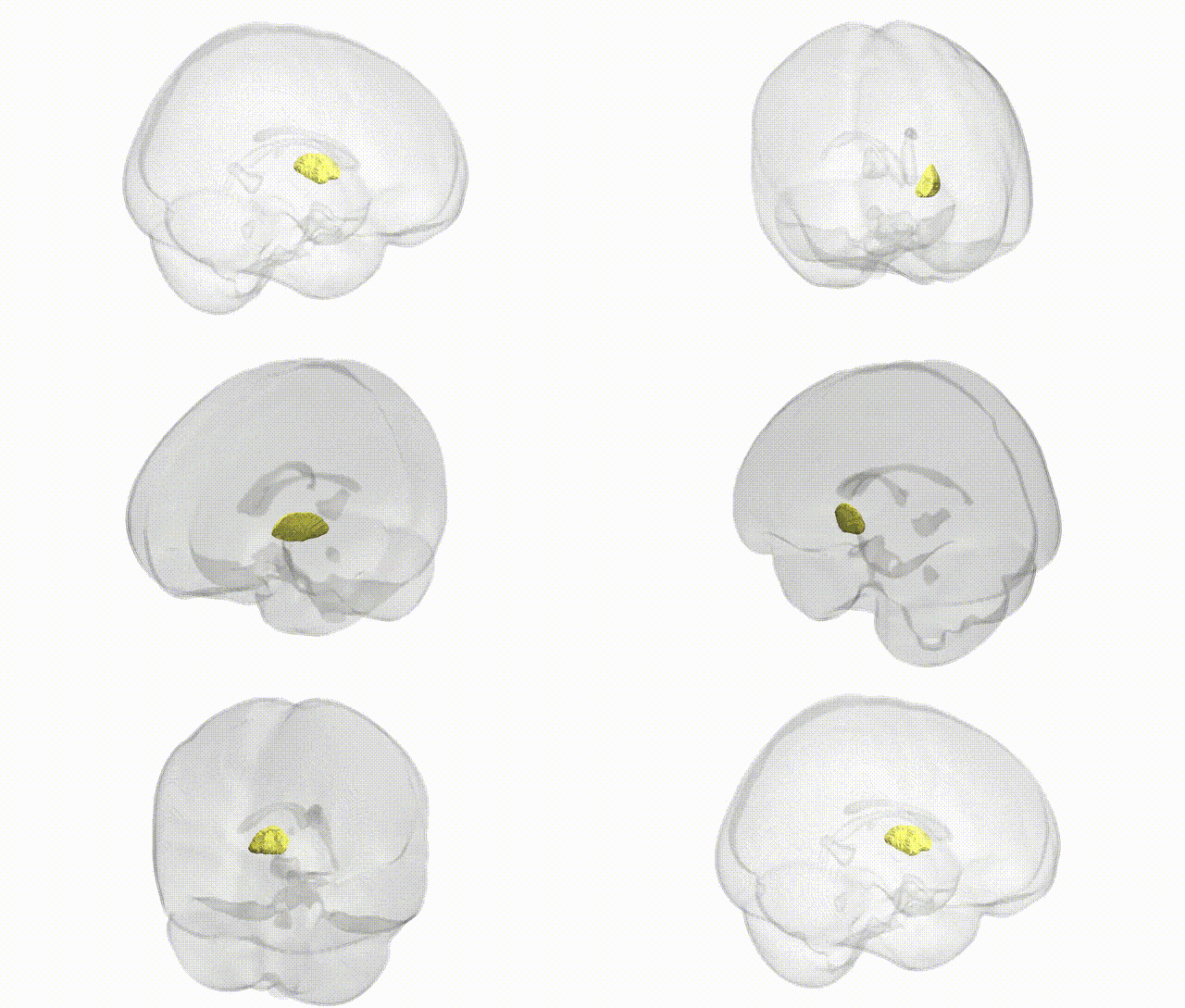
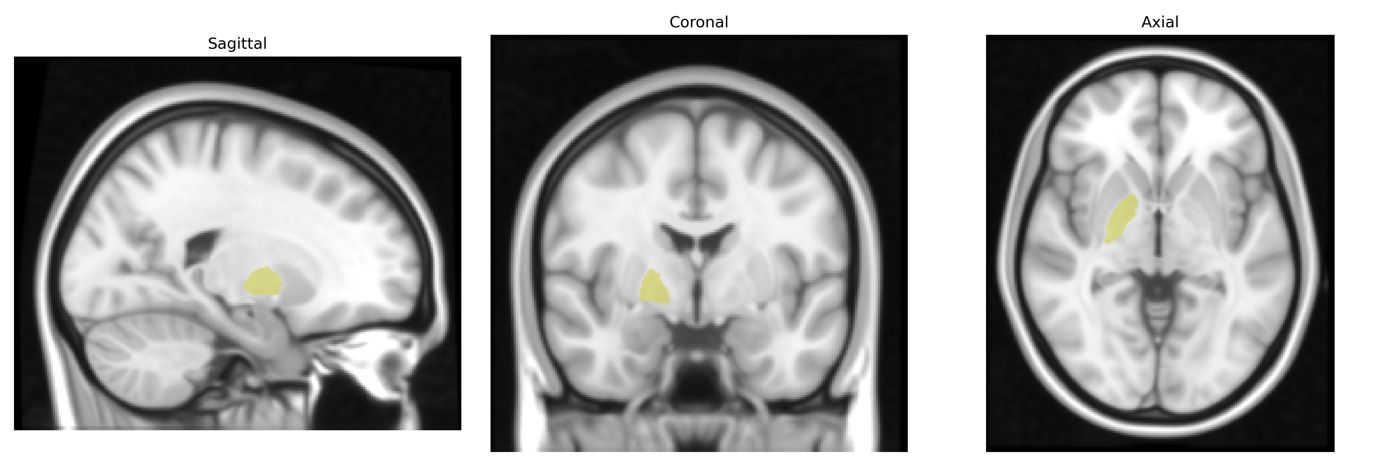
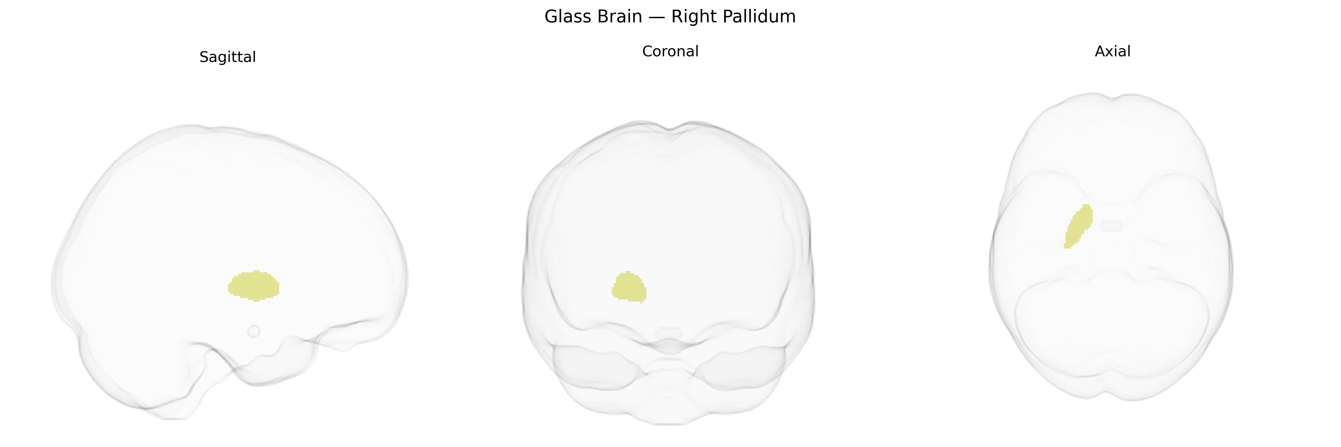
The bilateral right pallidum, as defined in the Harvard-Oxford subcortical structural atlas, corresponds to the right globus pallidus, a major component of the basal ganglia located medial to the putamen and lateral to the internal capsule. It is subdivided into internal (GPi) and external (GPe) segments, which receive dense input from the striatum and project to thalamic and brainstem nuclei via largely GABAergic (inhibitory) pathways. Functionally, the right pallidum participates in the regulation of voluntary movement, muscle tone, and motor learning, and also contributes to cognitive and limbic circuits involved in habit formation and reward-related behaviors. Pathophysiological involvement of this structure is prominent in movement disorders such as Parkinson’s disease and dystonia, where altered pallidal activity disrupts normal thalamo-cortical signaling. Globus pallidus
The right pallidum, part of the basal ganglia circuitry and often examined via the Harvard-Oxford subcortical atlas, shows robust genetic influences on its volume and function, with SNP-based heritability estimates from large MRI-GWAS (e.g., ENIGMA, UK Biobank) implicating genes involved in neurodevelopment, synaptic plasticity, and dopaminergic signaling (such as DRD2, CNTNAP2, and MAPT in broader basal ganglia analyses). GWAS of subcortical volumes have linked variants near KTN1, DCC, and BDNF to pallidal structure, while polygenic risk for schizophrenia, bipolar disorder, and major depressive disorder correlates with alterations in pallidal volume and connectivity. Parkinson’s disease and dystonia risk loci, including variants in genes like GBA, LRRK2, and TOR1A, have been associated with functional and structural changes affecting the globus pallidus, reflecting its central role in motor control and basal ganglia-thalamo-cortical loops. Additional associations include genetic overlaps with obsessive-compulsive disorder, Tourette syndrome, and addiction-related traits, where dopaminergic and glutamatergic genes modulate pallidal circuits, as well as cognitive and personality-related GWAS findings linking polygenic scores for intelligence, educational attainment, and neuroticism to pallidal morphology and activity, although many associations are shared across basal ganglia structures and are not exclusively specific to the right pallidum.
Overview generated by GPT-4o (2026).
Region ID: 18
Hemisphere: bilateral
Atlas: HarvardOxford sub maxprob thr25 1mm
Full Quality Version: Download MP4
Full Quality Version: Download MP4
Wali Sidiqyar*, Gaurav Rudravaram*, Elyssa M. McMaster, Trent M. Schwartz, Adam M. Saunders, Kurt G. Schilling, Bennett A. Landman "Introducing SPINS: A Shared Public Visualization Library of Neuroanatomical Structures." Medical Imaging with Deep Learning- short paper
This resource is licensed under CC0 1.0 Universal (Public Domain).