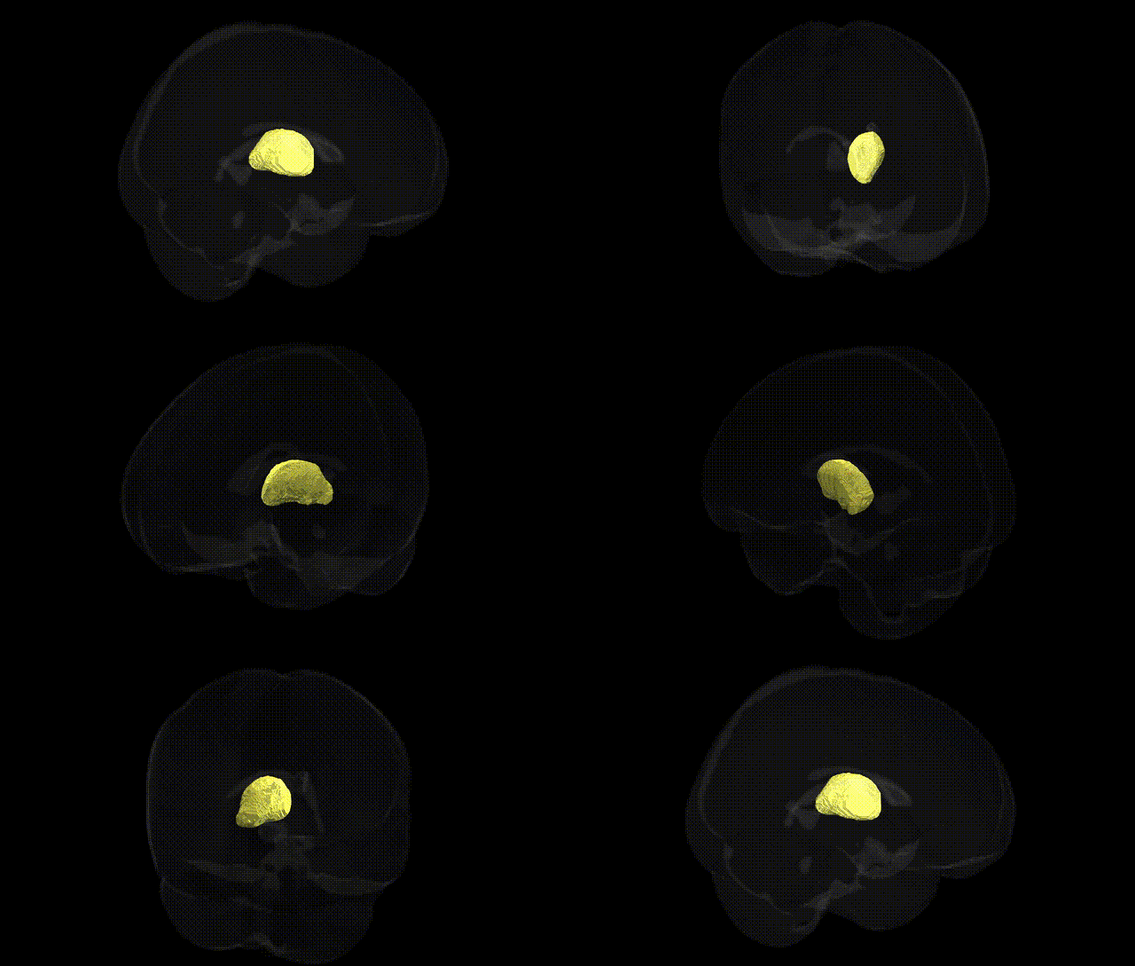
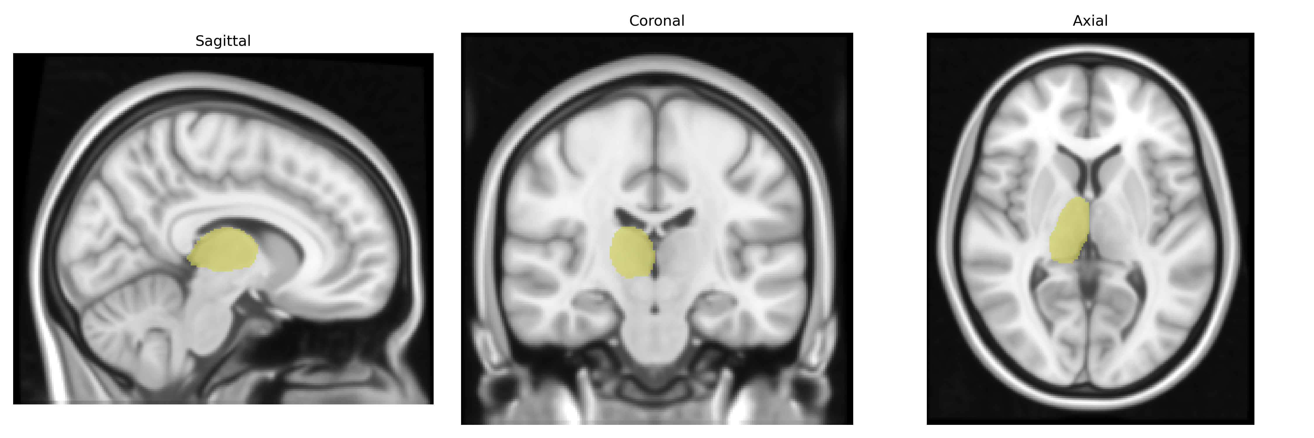
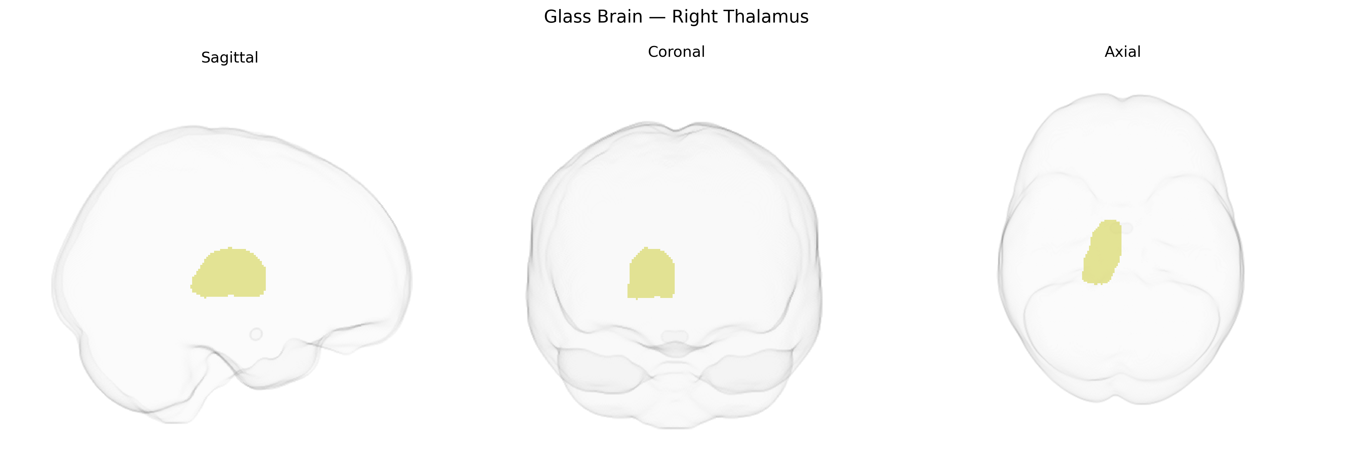
The bilateral Right Thalamus, as defined in the Harvard-Oxford Subcortical Atlas, comprises the right-sided portion of the thalamic complex, a paired diencephalic structure situated superior to the brainstem and medial to the internal capsule. It consists of multiple nuclei that serve as major relay and integrative stations, transmitting and modulating sensory (excluding olfactory), motor, limbic, and associative information between subcortical structures and the cerebral cortex. Functionally, the thalamus is critical for conscious perception, arousal, attentional gating, and the coordination of cortical activity patterns, with specific thalamic nuclei contributing to pain processing, visual and auditory pathways, motor control via cerebellar and basal ganglia loops, and higher cognitive processes. In the Harvard-Oxford atlas, the “Right Thalamus” label captures this heterogeneous nuclear region on the right hemisphere as a single anatomical unit, reflecting its role as a central hub in distributed brain networks. Thalamus
Genetic associations involving the bilateral right thalamus from large-scale neuroimaging GWAS (e.g., ENIGMA, UK Biobank) indicate that thalamic volume, including right-sided nuclei, is a highly polygenic trait with contributions from multiple loci implicated in neurodevelopment and synaptic function, including variants near genes such as BDNF, DLG2, SLC39A8, and others involved in neuronal signaling and brain morphogenesis. Thalamic structure has been linked to common psychiatric and neurological disorders—schizophrenia, bipolar disorder, major depressive disorder, autism spectrum disorder, attention-deficit/hyperactivity disorder, and Alzheimer’s disease—through both case-control imaging genetics and GWAS that show shared genetic architectures between thalamic volume and disease risk, particularly via pathways related to glutamatergic signaling, calcium channels, and immune regulation (e.g., complement system). Polygenic risk scores for schizophrenia and other psychotic disorders have been associated with altered thalamic morphology, including asymmetries that can involve the right thalamus, and several cognitive traits—general intelligence, processing speed, working memory—show genetic correlations with thalamic volume. Although most GWAS treat thalamus as a bilateral or total-volume phenotype rather than isolating the right side specifically, the genetic variants influencing global thalamic morphology are thought to contribute similarly to right-sided subregional anatomy as defined in the Harvard-Oxford subcortical atlas.
Overview generated by GPT-4o (2026).
Region ID: 15
Hemisphere: bilateral
Atlas: HarvardOxford sub maxprob thr25 1mm
Full Quality Version: Download MP4
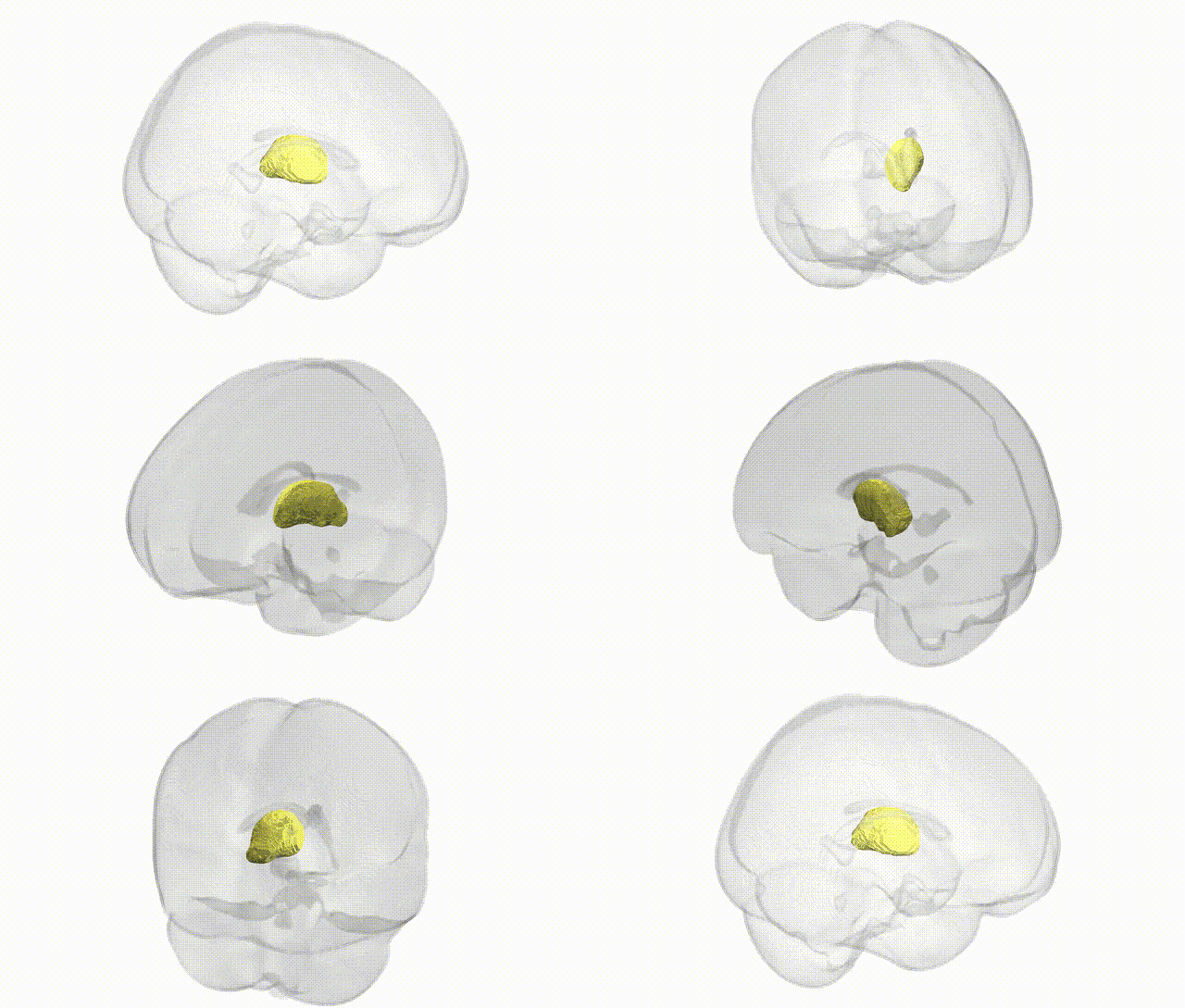
Full Quality Version: Download MP4
Wali Sidiqyar*, Gaurav Rudravaram*, Elyssa M. McMaster, Trent M. Schwartz, Adam M. Saunders, Kurt G. Schilling, Bennett A. Landman "Introducing SPINS: A Shared Public Visualization Library of Neuroanatomical Structures." Medical Imaging with Deep Learning- short paper
This resource is licensed under CC0 1.0 Universal (Public Domain).