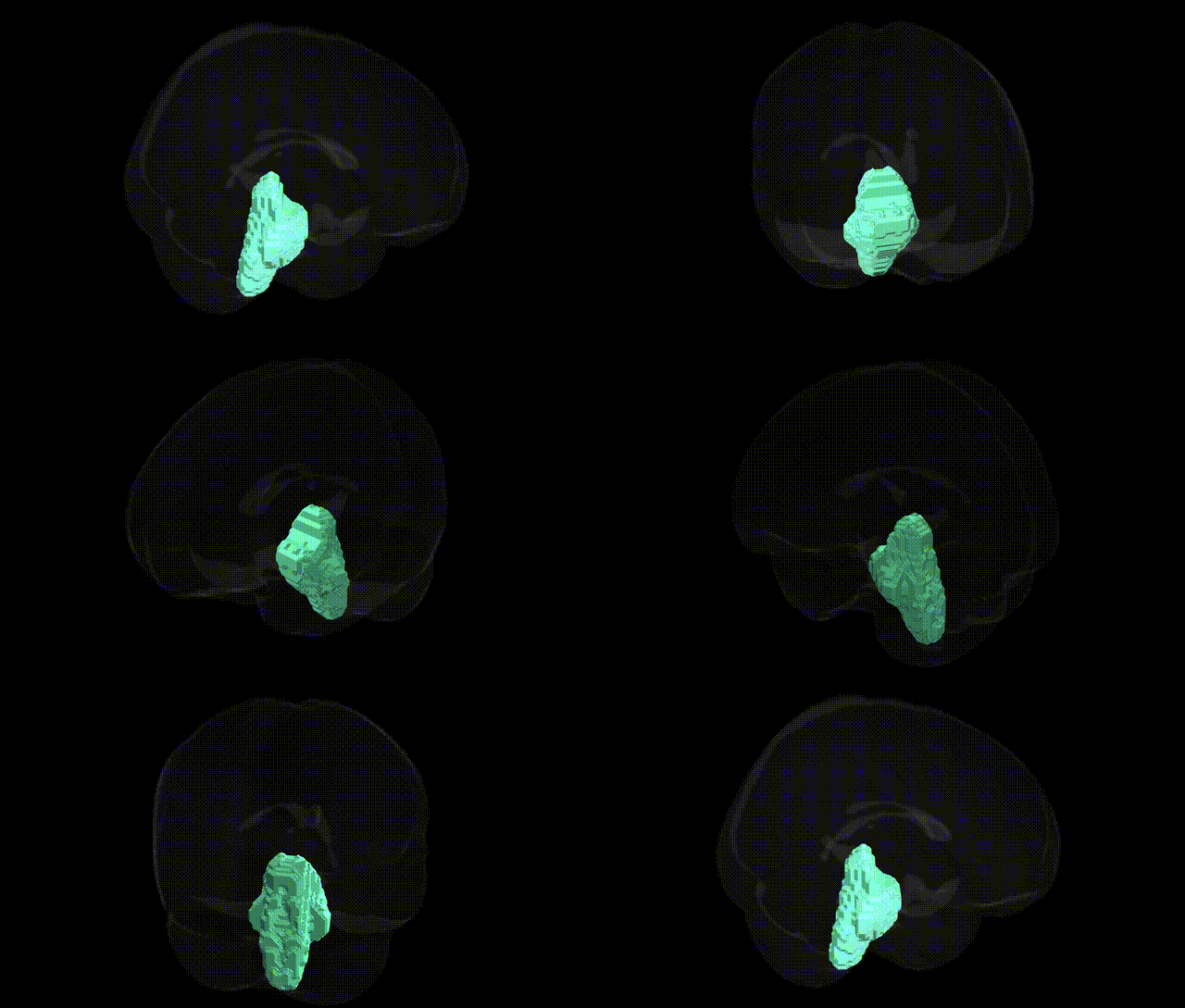
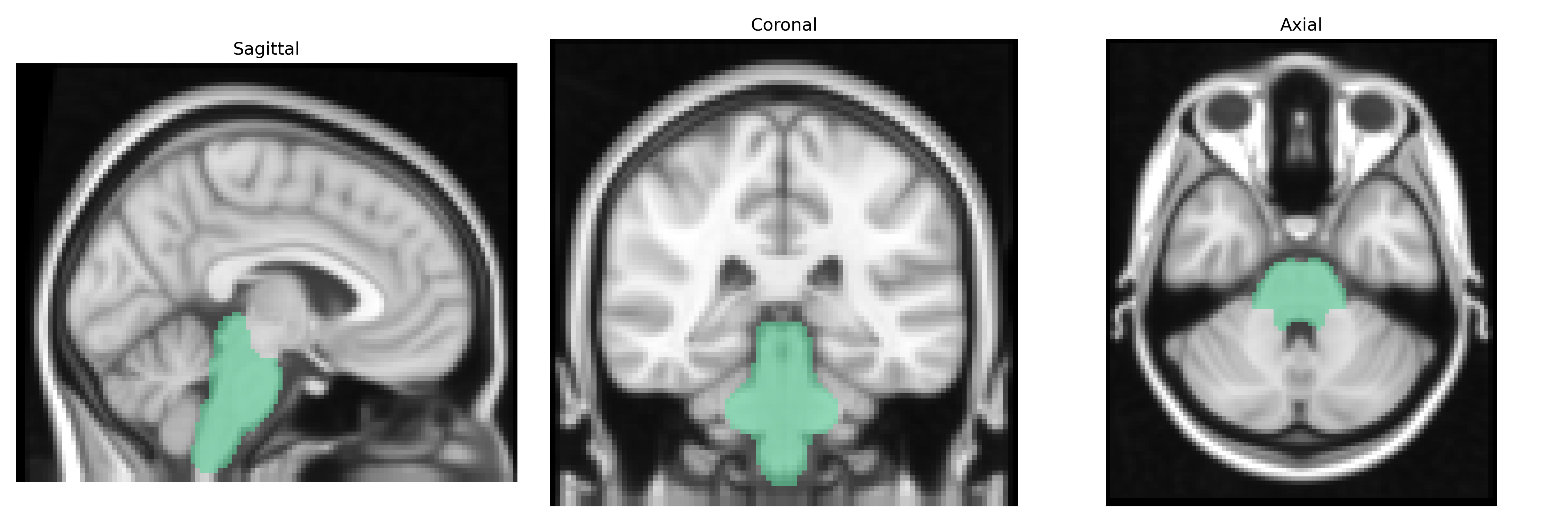
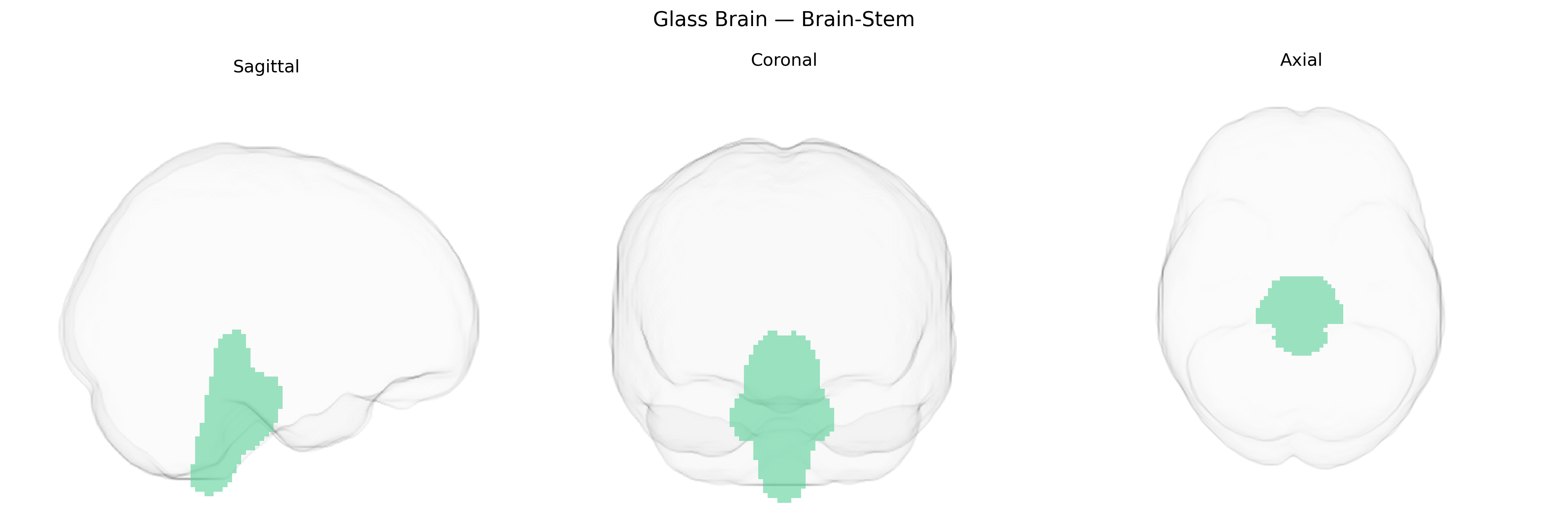
The bilateral brain-stem region in the Harvard-Oxford subcortical maxprob thr25 2mm atlas corresponds to the compact, centrally located structure at the base of the brain that connects the cerebrum and cerebellum with the spinal cord. It includes the midbrain, pons, and medulla oblongata, which collectively contain critical ascending sensory and descending motor pathways, cranial nerve nuclei, and autonomic centers essential for respiration, cardiovascular regulation, arousal, sleep–wake cycles, and basic reflexes such as swallowing and coughing. Functionally, the brain-stem integrates signals between higher cortical areas and peripheral systems, supports posture and locomotion via reticulospinal and vestibulospinal tracts, and maintains vital homeostatic control through interactions with the hypothalamus and other subcortical structures. Brainstem
The bilateral brainstem, as defined in the Harvard–Oxford subcortical atlas, has been implicated in genetic studies largely through its roles in autonomic, motor, pain, and affective regulation rather than through highly region-specific loci. Imaging genetics and GWAS of brain structure show that brainstem volume and microstructure are moderately heritable, with polygenic influences from common variants associated with general brain size (e.g., variants near HMGA2 and IGF1), neurodevelopmental pathways, and myelination. Genetic risk for neurodegenerative disorders such as Parkinson’s disease (e.g., SNCA, LRRK2, GBA), multiple sclerosis (HLA region and immune-related loci), and Alzheimer’s disease (APOE and other GWAS hits) has been linked to brainstem involvement in neuroimaging or neuropathology, particularly in structures like the substantia nigra, locus coeruleus, and dorsal raphe that lie within or adjacent to the atlas-defined brainstem. GWAS for migraine (e.g., TRPM8, LRP1, and other pain/modulation-related loci) and chronic pain, sleep traits, and autonomic dysfunction also indirectly implicate brainstem circuits, given its central role in nociceptive processing and arousal. Psychiatric genetics, including polygenic risk scores for major depressive disorder, anxiety, and PTSD, have been associated with functional and structural variation in brainstem monoaminergic nuclei, although individual loci are typically linked to neurotransmitter and synaptic genes (e.g., SLC6A4, MAOA, BDNF) rather than to the brainstem as a discrete anatomical target. Overall, genetic associations with the atlas-defined bilateral brainstem reflect distributed polygenic effects on neurodevelopment, neurotransmission, and neurodegeneration, with most findings inferred from system-level imaging and clinical phenotypes rather than brainstem-specific GWAS signals.
Overview generated by GPT-4o (2026).
Region ID: 8
Hemisphere: bilateral
Atlas: HarvardOxford sub maxprob thr25 2mm
Full Quality Version: Download MP4
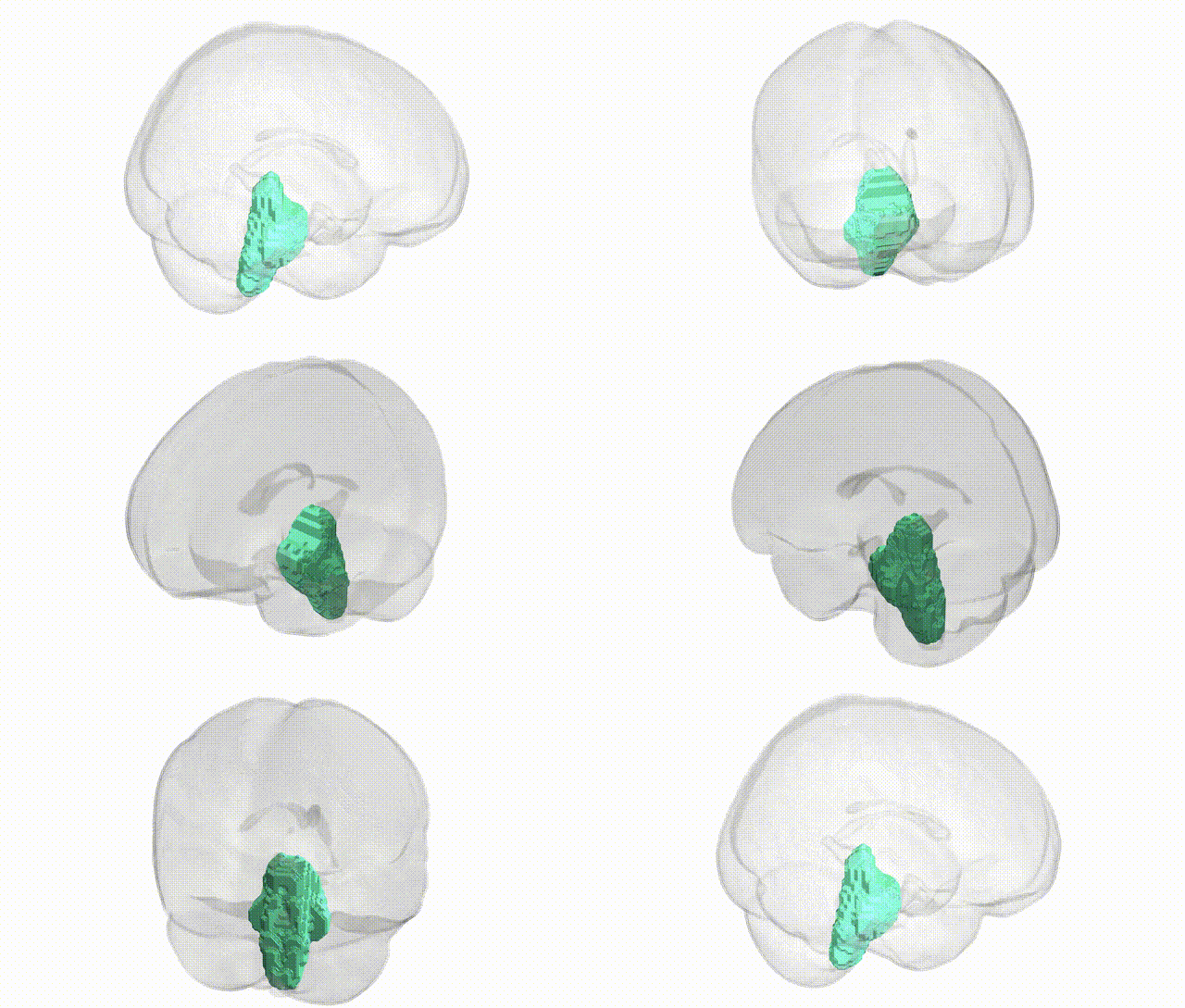
Full Quality Version: Download MP4
Wali Sidiqyar*, Gaurav Rudravaram*, Elyssa M. McMaster, Trent M. Schwartz, Adam M. Saunders, Kurt G. Schilling, Bennett A. Landman "Introducing SPINS: A Shared Public Visualization Library of Neuroanatomical Structures." Medical Imaging with Deep Learning- short paper
This resource is licensed under CC0 1.0 Universal (Public Domain).