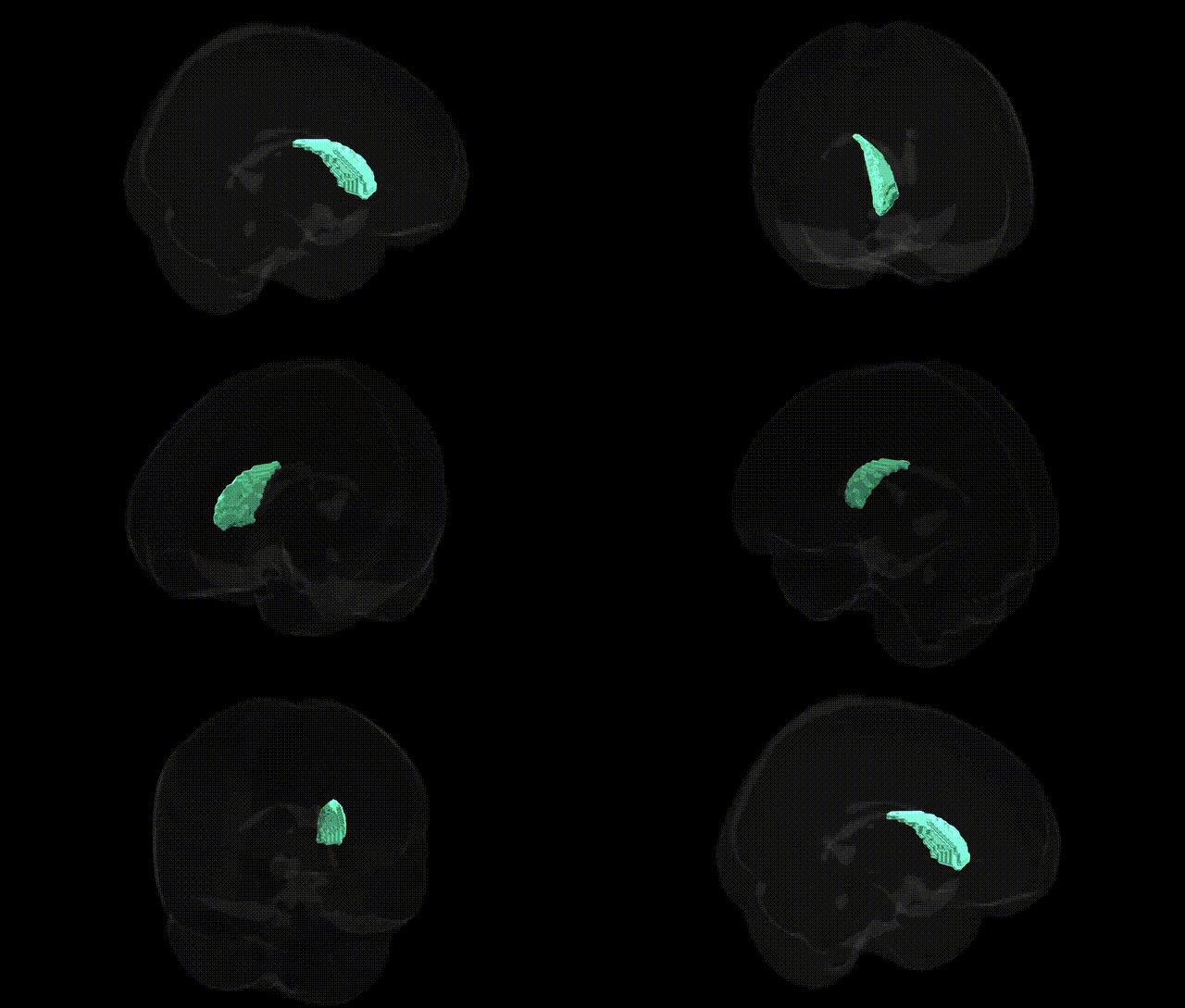
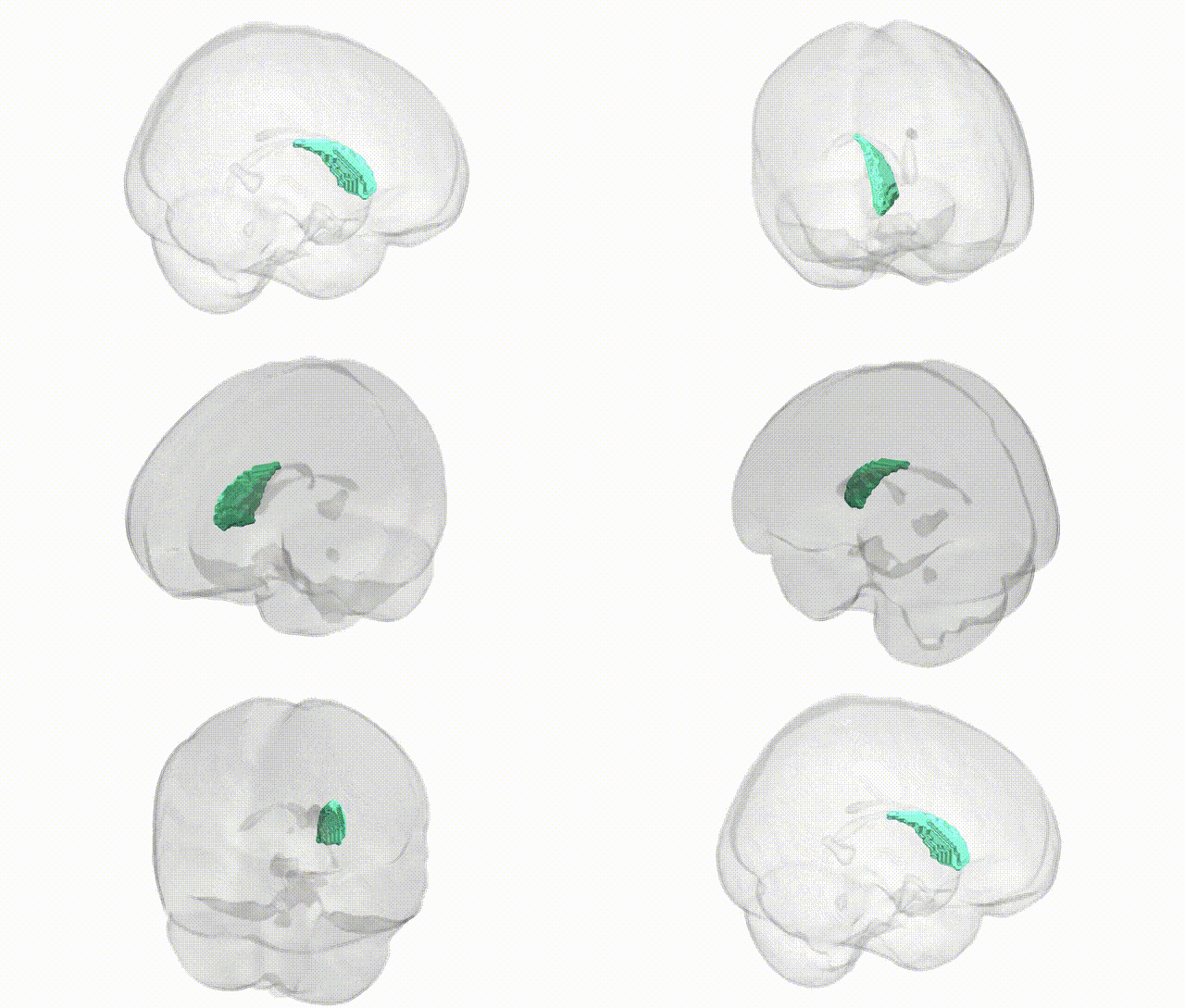
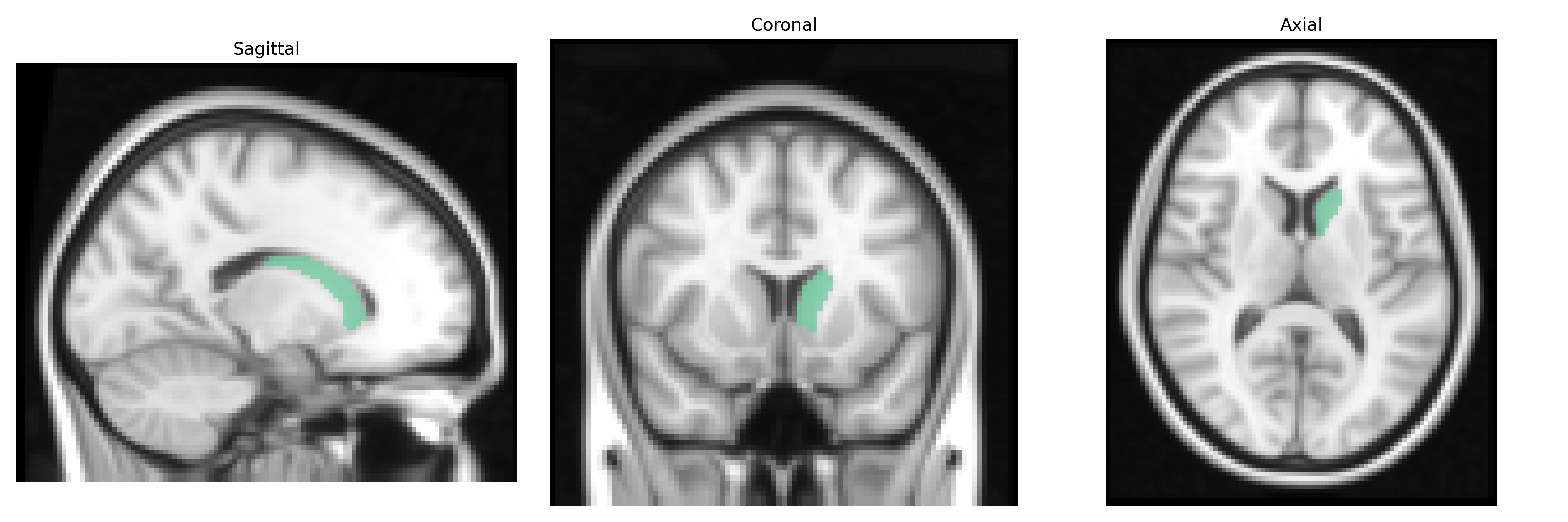
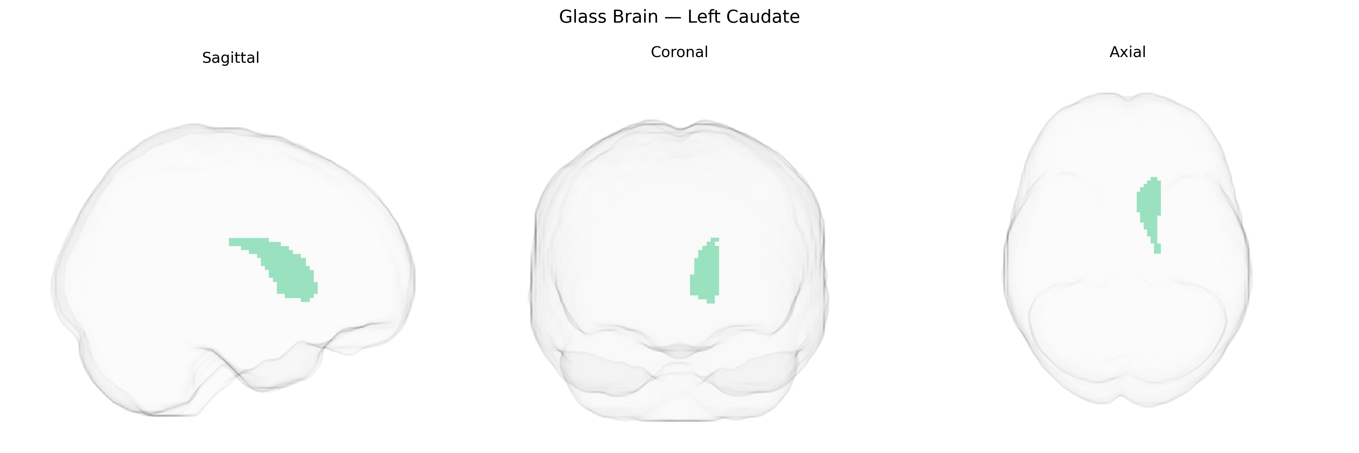
The bilateral Left Caudate, as defined in the Harvard-Oxford Subcortical Structural Atlas (sub maxprob thr25, 2 mm), refers to the left-sided component of the caudate nucleus, a C-shaped gray matter structure of the dorsal striatum situated deep within the basal ganglia and aligned along the lateral ventricles. It is cytoarchitectonically and functionally divided into head, body, and tail, with dense inputs from the cerebral cortex (especially prefrontal and association areas) and primary outputs via the globus pallidus and substantia nigra, thereby participating in cortico-striato-thalamo-cortical loops. The left caudate nucleus plays key roles in motor control, action selection, procedural learning, reward processing, habit formation, and higher-order cognitive and executive functions, and is implicated in multiple neurological and psychiatric conditions, including Parkinson’s disease, Huntington’s disease, obsessive-compulsive disorder, and attention-deficit/hyperactivity disorder. Caudate nucleus
The bilateral left caudate, as defined in the Harvard-Oxford subcortical atlas, has been repeatedly implicated in genetic studies of neuropsychiatric and cognitive traits, particularly through GWAS of brain structure and function. Variants in genes involved in dopamine signaling (e.g., DRD2, COMT) and synaptic plasticity (e.g., BDNF) have been associated with caudate volume and activity, consistent with the region’s role in reward processing, habit formation, and executive control. Large-scale imaging genetics consortia such as ENIGMA and UK Biobank–based GWAS have identified multiple loci influencing caudate volume, including variants near HMGA2, KTN1, and genes related to neurodevelopment, neuronal migration, and cytoskeletal organization, some of which show pleiotropic associations with general cognitive ability, educational attainment, and intracranial volume. The caudate has also emerged in genetic studies of psychiatric disorders: polygenic risk for schizophrenia, bipolar disorder, and major depressive disorder has been linked to altered caudate volume and connectivity, while loci associated with obsessive-compulsive disorder, attention-deficit/hyperactivity disorder, and Tourette syndrome often map to pathways implicated in cortico-striato-thalamo-cortical circuits that prominently involve the caudate. Furthermore, GWAS of Parkinson’s disease, Huntington’s disease (where the caudate is a primary site of atrophy), and substance use disorders highlight genes affecting striatal medium spiny neurons and dopaminergic modulation, supporting a genetically influenced vulnerability of the caudate to motor, compulsive, and addictive phenotypes.
Overview generated by GPT-4o (2026).
Region ID: 5
Hemisphere: bilateral
Atlas: HarvardOxford sub maxprob thr25 2mm
Full Quality Version: Download MP4
Full Quality Version: Download MP4
Wali Sidiqyar*, Gaurav Rudravaram*, Elyssa M. McMaster, Trent M. Schwartz, Adam M. Saunders, Kurt G. Schilling, Bennett A. Landman "Introducing SPINS: A Shared Public Visualization Library of Neuroanatomical Structures." Medical Imaging with Deep Learning- short paper
This resource is licensed under CC0 1.0 Universal (Public Domain).