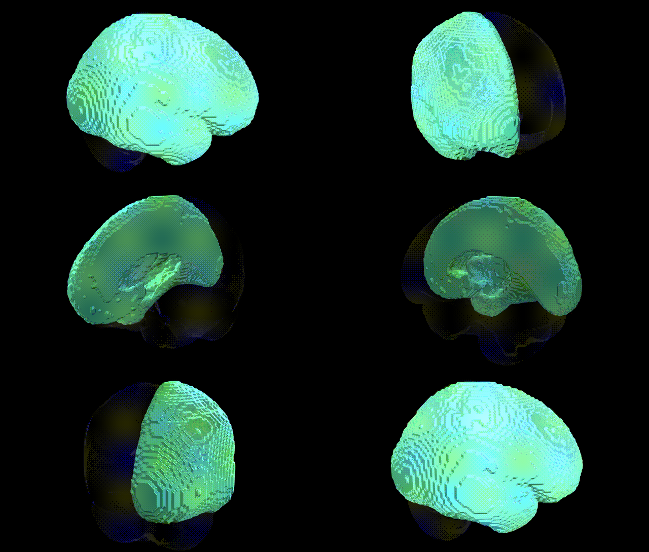
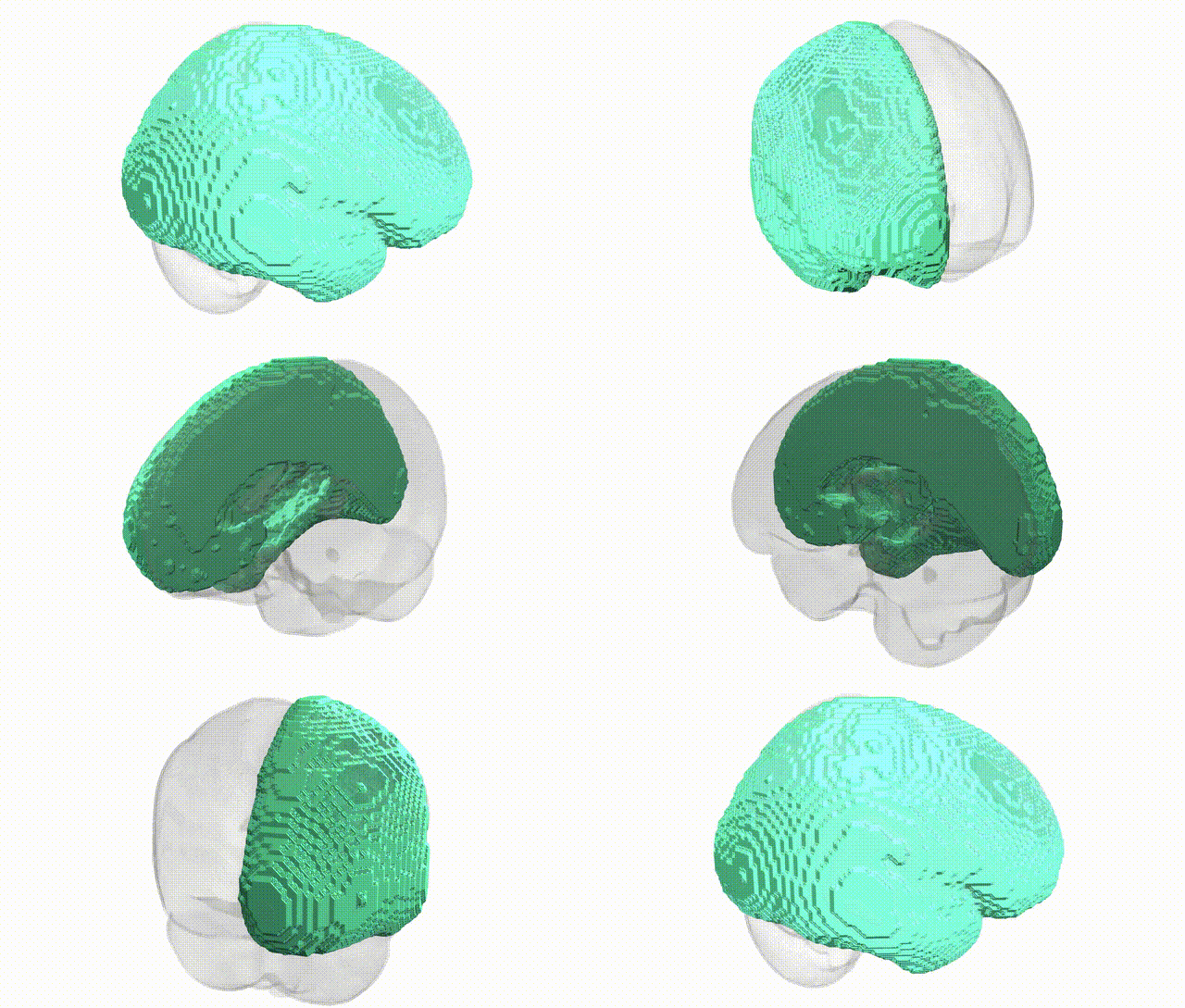
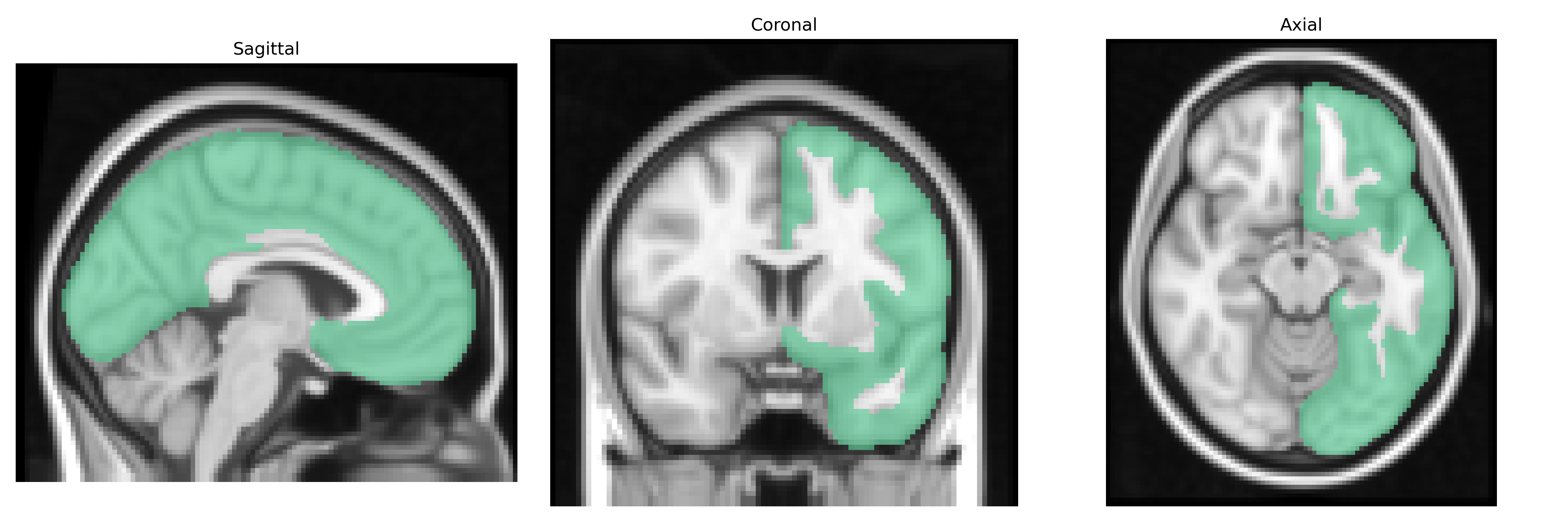
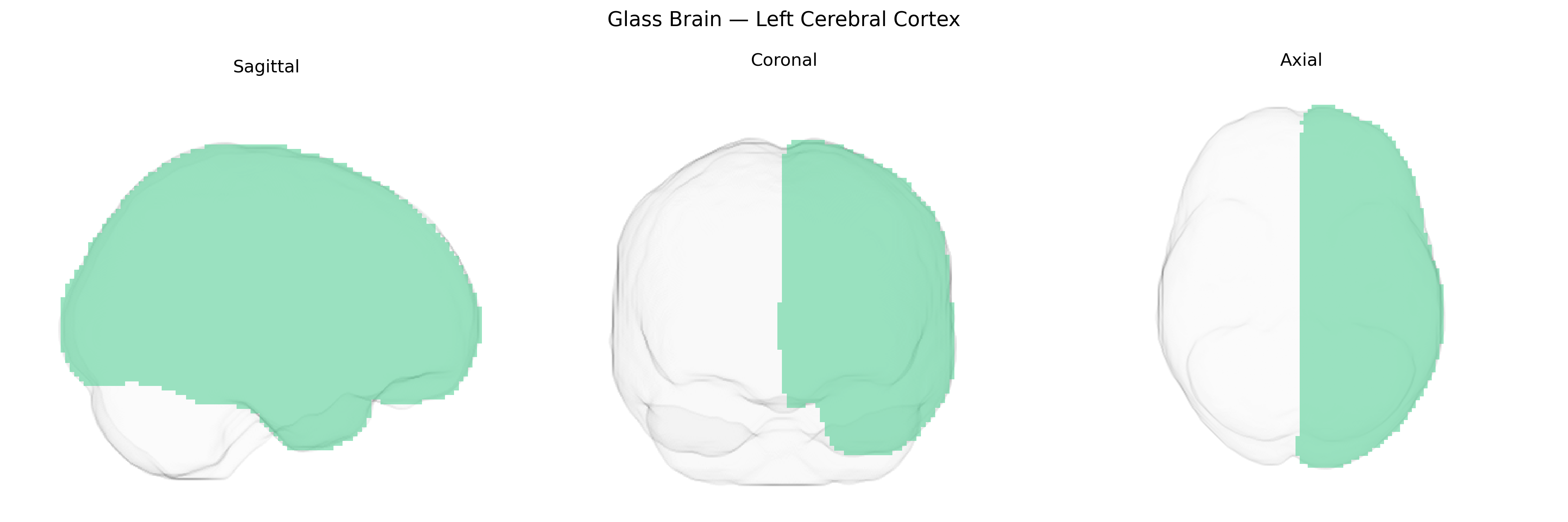
The bilateral Left Cerebral Cortex in the Harvard-Oxford subcortical maximum probability atlas (thr25, 2 mm) refers to the left-sided component of the cerebral cortex, encompassing the outer laminar sheet of gray matter that covers the left cerebral hemisphere and is involved in higher-order cognitive, sensory, motor, and associative processing. Structurally, it consists of six-layered neocortex (and small allocortical regions), organized into distinct functional areas supporting language (typically dominant in the left hemisphere), voluntary motor control, somatosensory integration, perception, memory, and executive functions. The cortex is highly interconnected via intracortical and subcortical pathways, with columnar and laminar organization underlying complex information processing and plasticity. There is no direct link for “Left Cerebral Cortex,” but it is part of the broader cerebral cortex structure: Cerebral cortex.
The bilateral left cerebral cortex as defined in the Harvard–Oxford subcortical maximum probability atlas is a gross anatomical label rather than a functionally specific region, so genetic associations typically arise from large-scale brain imaging genetics and GWAS of cortical structure and related disorders rather than this atlas label itself. GWAS of cortical thickness, surface area, and volume (e.g., ENIGMA, UK Biobank) have identified numerous loci affecting global and regional left-hemisphere cortical measures, with robust signals near genes such as TBR1, CENPW, RSPO3, HMGA2, and DACT1, implicating pathways in neurodevelopment, Wnt signaling, and cell proliferation. Hemispheric and regional asymmetries of the left cortex—particularly in language-related areas—show heritable variation with loci near LRRTM4, PCSK6, and DCDC2 and overlap with genetic risk for developmental dyslexia, specific language impairment, and schizophrenia. Global and lobar left cortical metrics are also influenced by polygenic risk for neuropsychiatric and cognitive traits, including schizophrenia, major depression, bipolar disorder, autism spectrum disorder, and educational attainment, with shared variants in synaptic, calcium signaling, and chromatin remodeling genes (e.g., CACNA1C, GRIN2A, MIR137 loci). In neurodegenerative disease, GWAS for cortical atrophy patterns and left-dominant temporal and parietal thinning in Alzheimer’s disease highlight APOE, BIN1, CLU, and CR1, linking classical AD risk loci to structural vulnerability of the left cortex. Overall, genetic influences on the bilateral left cerebral cortex reflect a diffuse, polygenic architecture that overlaps strongly with genes for general brain development, cognitive function, and risk for major psychiatric and neurodegenerative disorders, rather than a set of variants unique to this Harvard–Oxford atlas-defined region.
Overview generated by GPT-4o (2026).
Region ID: 2
Hemisphere: bilateral
Atlas: HarvardOxford sub maxprob thr25 2mm
Full Quality Version: Download MP4
Full Quality Version: Download MP4
Wali Sidiqyar*, Gaurav Rudravaram*, Elyssa M. McMaster, Trent M. Schwartz, Adam M. Saunders, Kurt G. Schilling, Bennett A. Landman "Introducing SPINS: A Shared Public Visualization Library of Neuroanatomical Structures." Medical Imaging with Deep Learning- short paper
This resource is licensed under CC0 1.0 Universal (Public Domain).