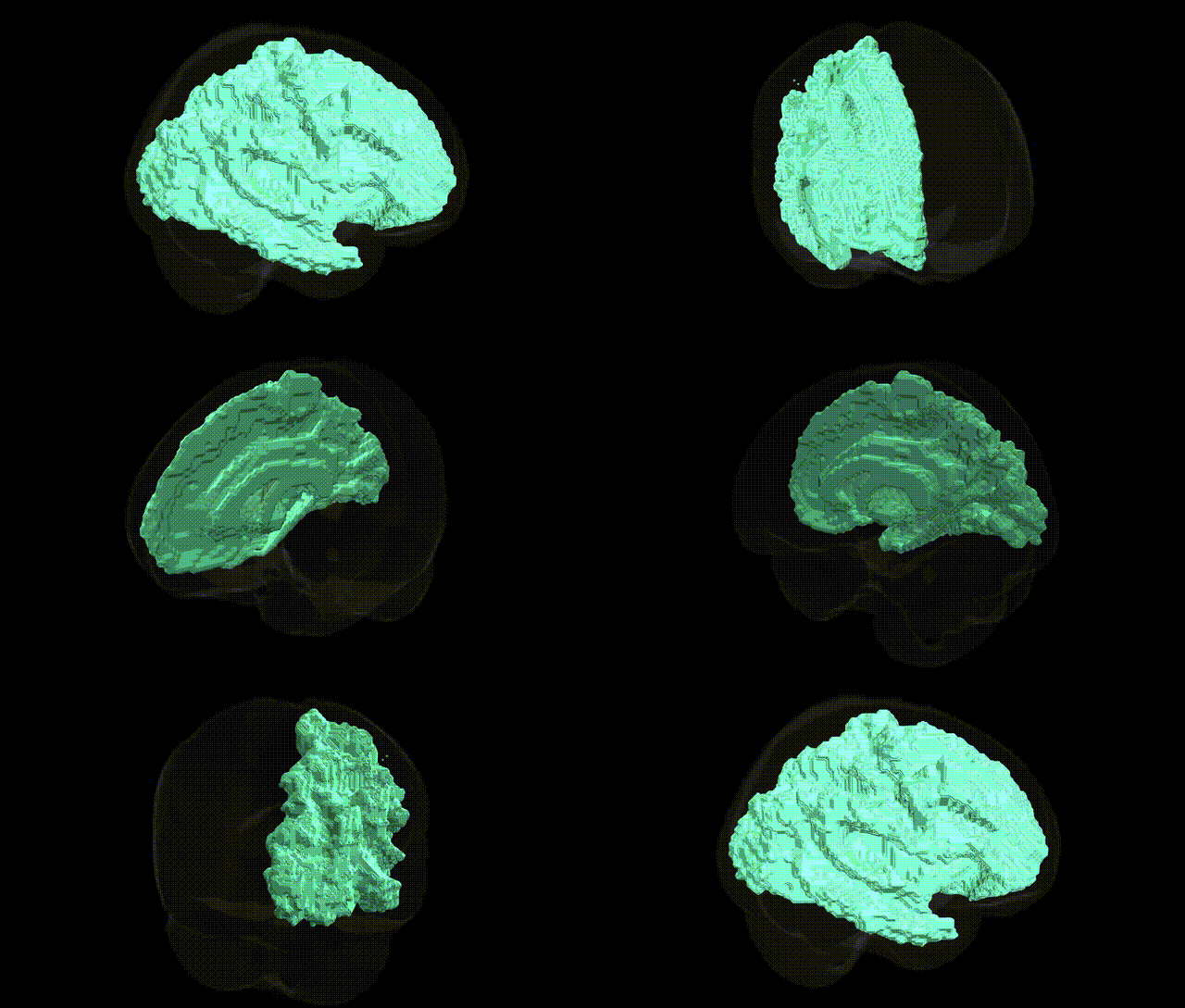
The bilateral left cerebral white matter comprises the myelinated axonal tracts underlying the cerebral cortex in the left hemisphere, forming the principal conduit for long-range communication among cortical regions and between cortex and subcortical structures. This tissue includes association fibers (linking cortical areas within the hemisphere), commissural fibers (primarily the portion of the corpus callosum projecting to the left), and projection fibers (connecting cortex with deep nuclei and brainstem), supporting functions such as language, motor control, sensory integration, and higher cognition by enabling rapid signal transmission. Structural integrity of left cerebral white matter is critical for efficient neural processing, and its disruption is implicated in conditions like multiple sclerosis, stroke, traumatic brain injury, and various neurodevelopmental and psychiatric disorders. There is no direct Wikipedia article for “Left Cerebral White Matter”; see the related entry on White matter.
Genetic associations specifically targeting the “bilateral Left Cerebral White Matter” label from the Harvard–Oxford atlas are not typically reported at that exact regional resolution, but numerous GWAS and imaging‑genetics studies have identified variants influencing global and lobar cerebral white matter microstructure, volume, and integrity that would encompass this region. Common variants in genes related to myelination and axonal integrity—such as BIN1, CLU, PICALM, APOE, SORL1, and other Alzheimer’s disease–associated loci—have been linked to widespread white matter changes, including reduced fractional anisotropy and increased white matter hyperintensities, which likely affect left cerebral white matter. Large ENIGMA consortium studies and UK Biobank GWAS have shown that polygenic risk for schizophrenia, bipolar disorder, major depression, autism spectrum disorder, and ADHD is associated with altered white matter microstructure across major tracts (e.g., corpus callosum, cingulum, superior longitudinal fasciculus) that pass through left cerebral white matter, implicating synaptic, neurodevelopmental, and immune-related genes (such as CACNA1C, GRIN2A, ZNF804A, NRG1, and MHC region variants). Additionally, GWAS of white matter hyperintensity burden and small-vessel disease have identified variants near COL4A2, HTRA1, FOXC1, and other vascular and extracellular matrix genes, linking genetic risk for hypertension, stroke, and cognitive decline to diffuse white matter damage, including in left hemispheric regions. Collectively, genetic influences on myelination, neurodevelopment, synaptic signaling, and vascular integrity contribute to structural variation and disease‑related pathology in the cerebral white matter, though current evidence is regional and tract-based rather than specific to the atlas-defined “bilateral Left Cerebral White Matter” parcel.
Overview generated by GPT-4o (2026).
Region ID: 1
Hemisphere: bilateral
Atlas: HarvardOxford sub maxprob thr25 2mm
Full Quality Version: Download MP4
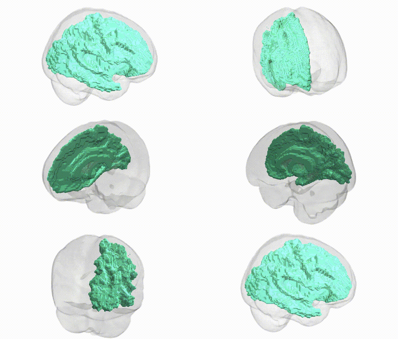
Full Quality Version: Download MP4
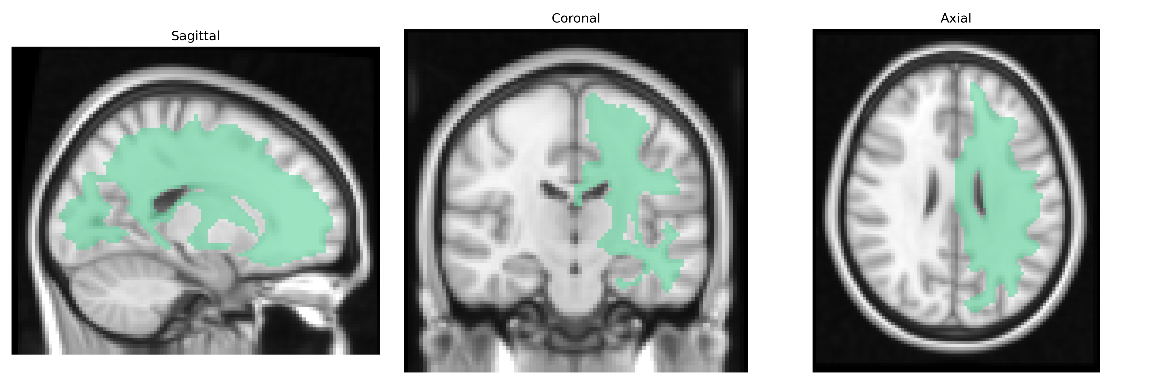
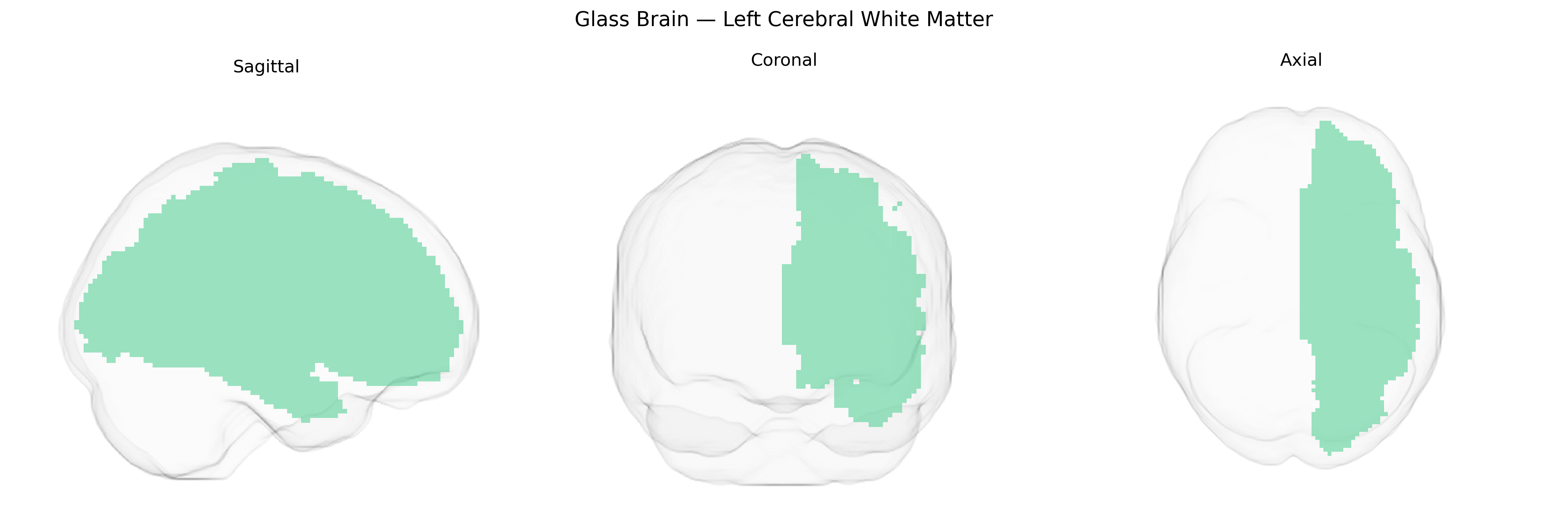
Wali Sidiqyar*, Gaurav Rudravaram*, Elyssa M. McMaster, Trent M. Schwartz, Adam M. Saunders, Kurt G. Schilling, Bennett A. Landman "Introducing SPINS: A Shared Public Visualization Library of Neuroanatomical Structures." Medical Imaging with Deep Learning- short paper
This resource is licensed under CC0 1.0 Universal (Public Domain).