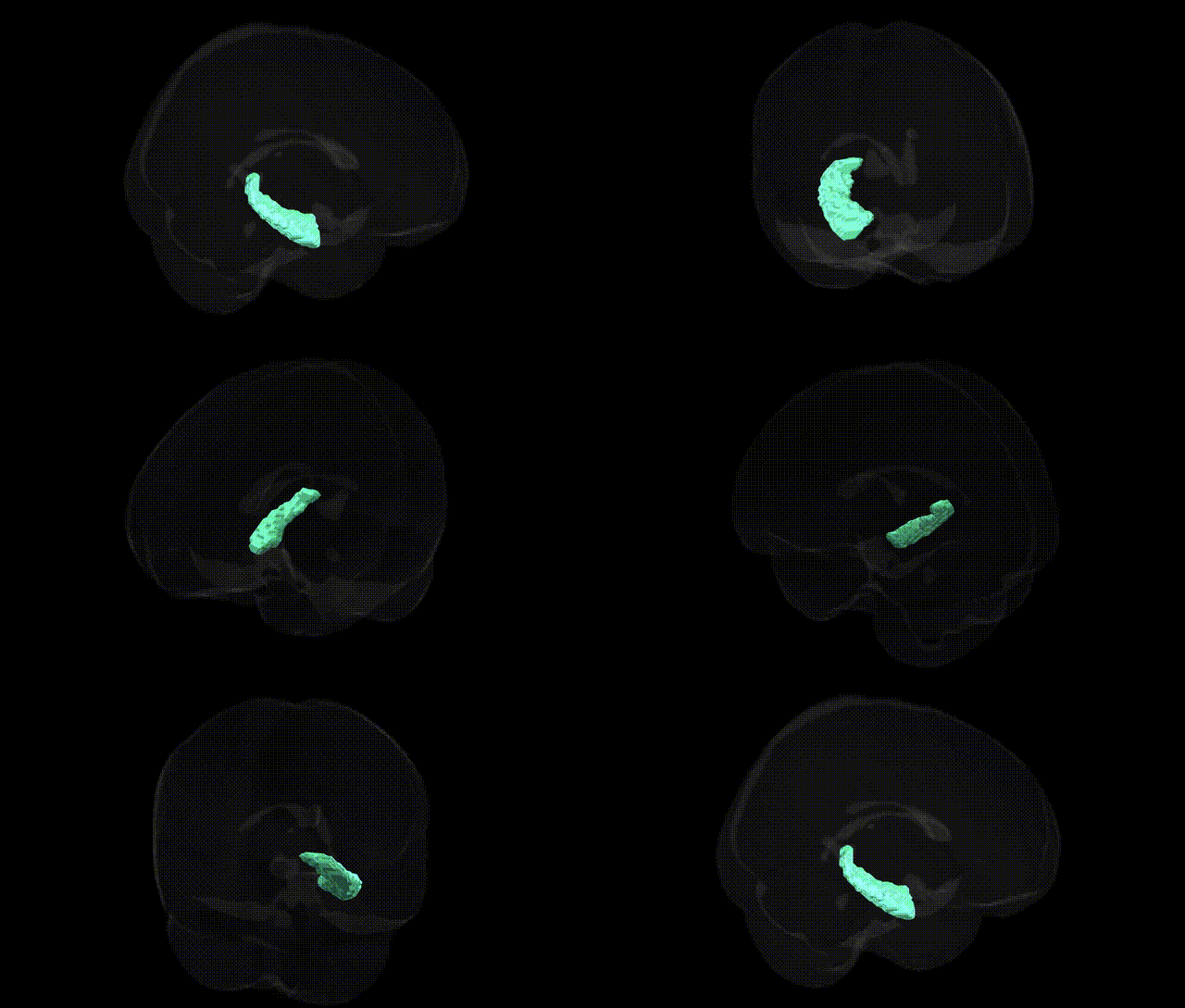
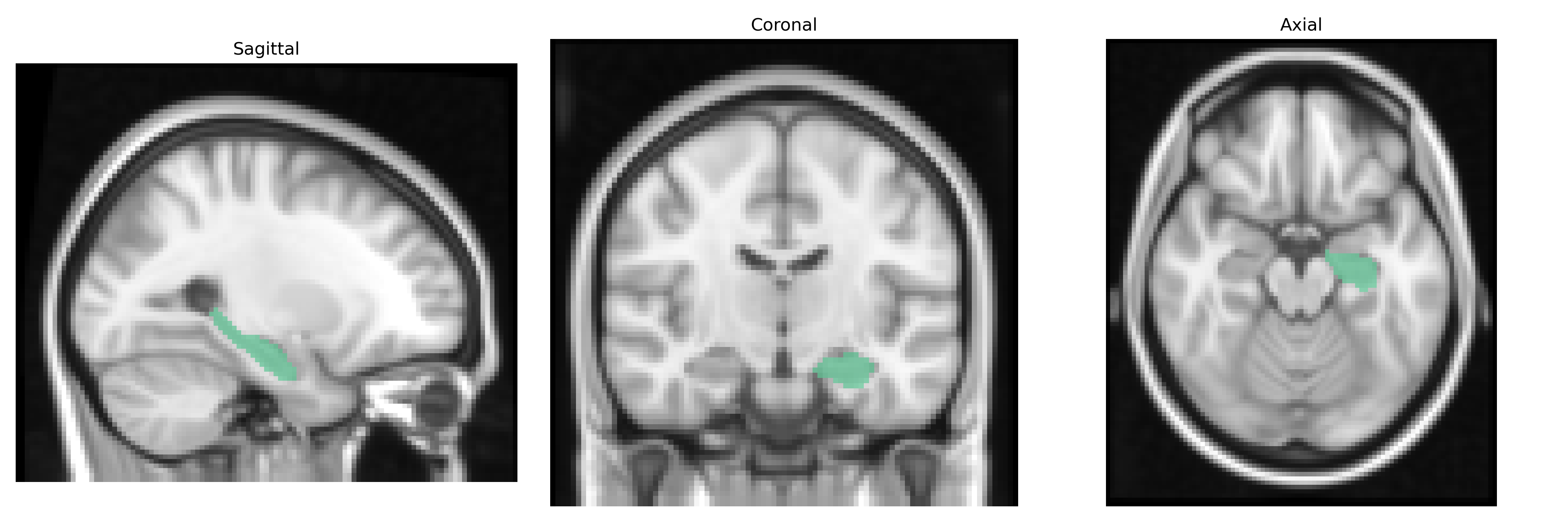
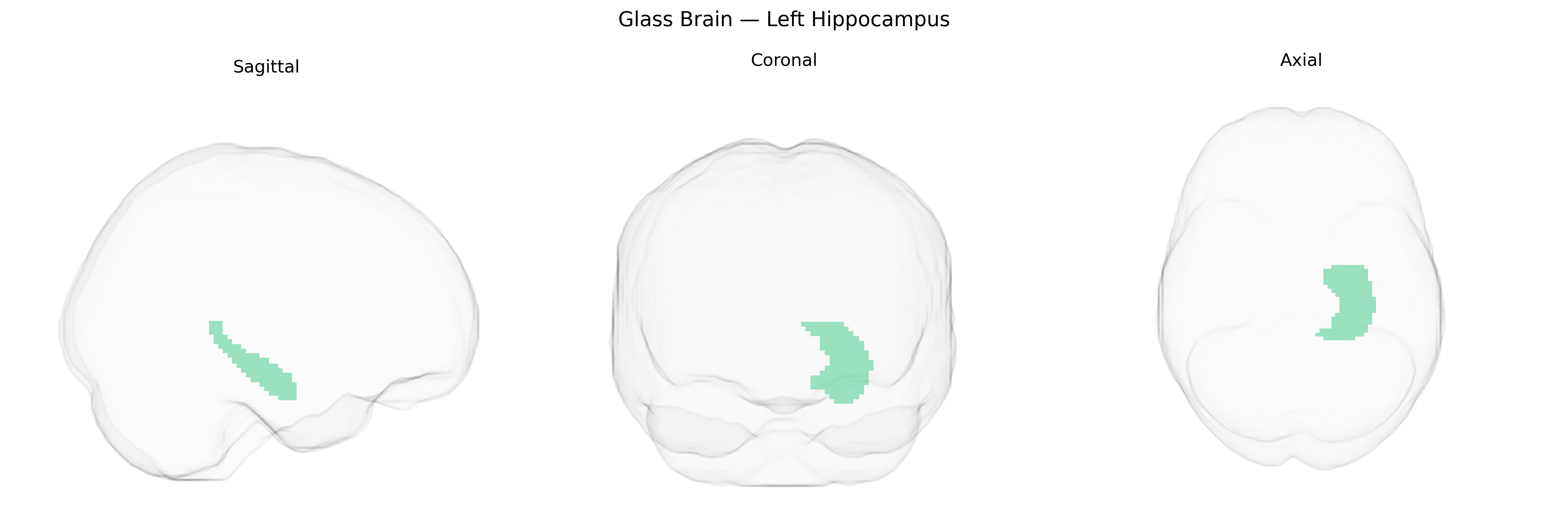
The bilateral Left Hippocampus, as delineated in the Harvard-Oxford subcortical maximum probability (thr25, 2 mm) atlas, corresponds to the left hippocampal formation, a medial temporal lobe structure crucial for episodic memory encoding, consolidation, and spatial navigation. It comprises subfields including the dentate gyrus, CA regions, and subiculum, and is heavily interconnected with the entorhinal cortex, parahippocampal gyrus, amygdala, and prefrontal areas, forming a core component of the limbic memory circuit. Functionally, the left hippocampus is particularly associated with verbal and autobiographical memory processes, contextual association learning, and the integration of multimodal sensory information into coherent, retrievable memory traces. Neurobiologically, it shows high synaptic plasticity and is vulnerable to pathological changes in conditions such as temporal lobe epilepsy, Alzheimer’s disease, and stress-related disorders. There is no direct Wikipedia article specifically for “Left Hippocampus”; a closely related and encompassing entry is Hippocampus.
The bilateral left hippocampus, as delineated in the Harvard-Oxford subcortical atlas, has been robustly implicated in genetic studies of memory, neurodegeneration, and psychiatric risk, largely via GWAS of hippocampal volume and function using MRI-based phenotypes. Common variants in genes related to neurodevelopment, synaptic plasticity, and neurodegenerative pathways—such as APOE (particularly the ε4 allele), CLU, PICALM, SORL1, and BIN1—have been associated with reduced hippocampal volume and increased risk for Alzheimer’s disease, while loci near genes like HMGA2, DPP4, and MSRB3 have been linked to general hippocampal volume and cognitive performance in large imaging-genetics cohorts. Polygenic risk scores for schizophrenia, major depressive disorder, and bipolar disorder show associations with structural and functional alterations in the left hippocampus, including reduced volume and abnormal connectivity, consistent with the region’s role in episodic memory, emotion regulation, and stress responsivity. Variants within BDNF (notably Val66Met), FKBP5, and other stress-related genes have been related to hippocampal volume and activity, particularly in the context of trauma exposure and PTSD, while genes involved in neurogenesis and glutamatergic signaling (e.g., GRIN2B, DISC1) have been linked to hippocampal-dependent learning and memory performance. Across disorders, convergent GWAS evidence indicates that genetically driven vulnerability impacting hippocampal integrity contributes to risk for Alzheimer’s disease, cognitive decline, recurrent major depression, schizophrenia, and stress-related psychopathology, with left-lateralized findings often emerging in memory and verbal learning paradigms, although most large-scale imaging GWAS treat bilateral or total hippocampal volume as the primary phenotype rather than strictly isolating the left subregion.
Overview generated by GPT-4o (2026).
Region ID: 9
Hemisphere: bilateral
Atlas: HarvardOxford sub maxprob thr25 2mm
Full Quality Version: Download MP4
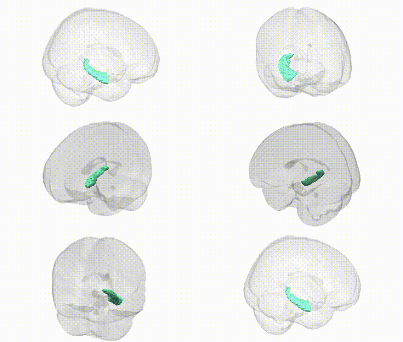
Full Quality Version: Download MP4
Wali Sidiqyar*, Gaurav Rudravaram*, Elyssa M. McMaster, Trent M. Schwartz, Adam M. Saunders, Kurt G. Schilling, Bennett A. Landman "Introducing SPINS: A Shared Public Visualization Library of Neuroanatomical Structures." Medical Imaging with Deep Learning- short paper
This resource is licensed under CC0 1.0 Universal (Public Domain).