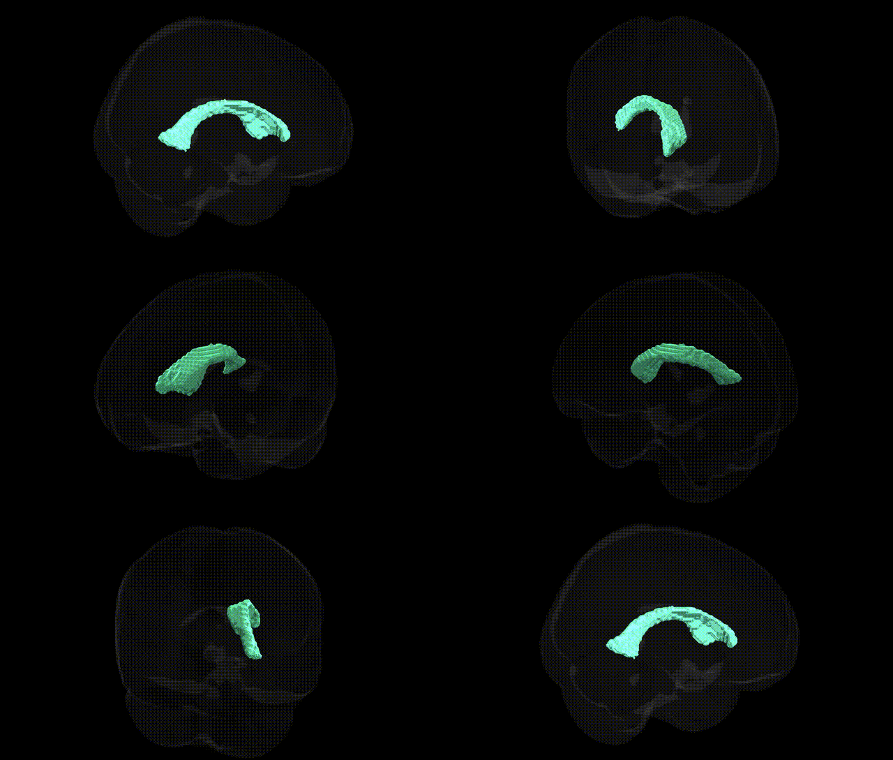
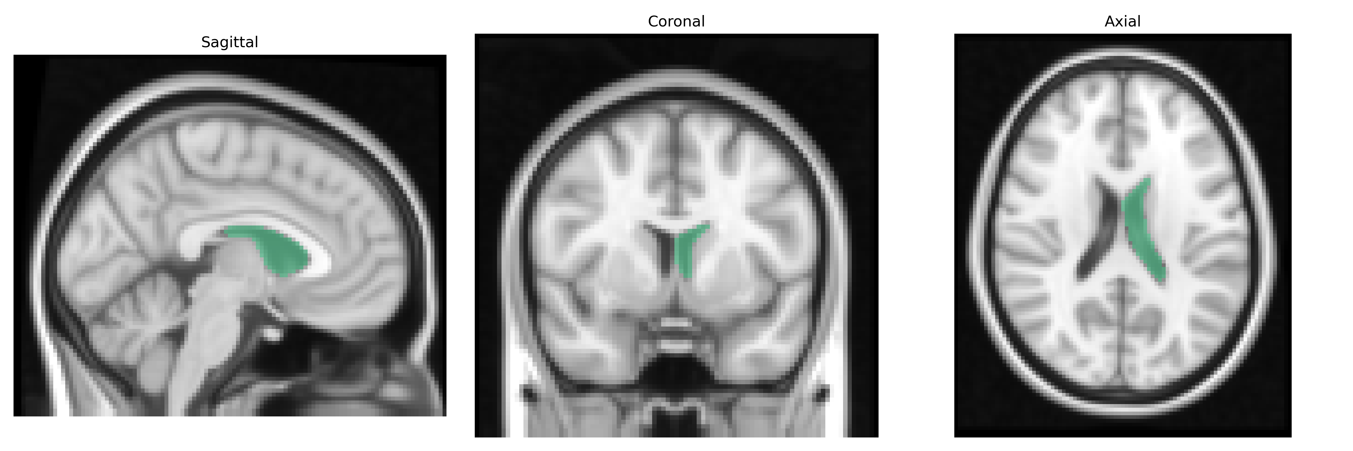
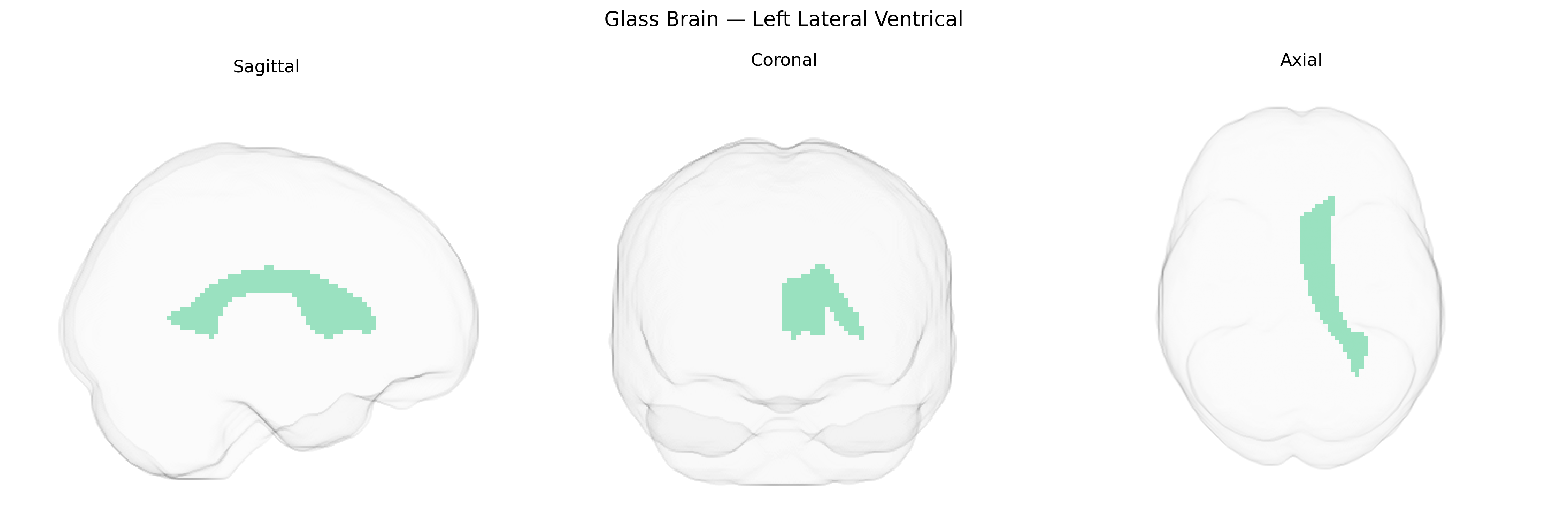
The bilateral left lateral ventricle, as defined in the Harvard-Oxford subcortical atlas, corresponds to the cerebrospinal fluid–filled cavity within the left cerebral hemisphere that lies deep to the cortex and adjacent to structures such as the caudate nucleus, thalamus, and corpus callosum. It consists of the frontal (anterior) horn, body, atrium (trigone), occipital (posterior) horn, and temporal (inferior) horn, each conforming to the shape and extent of the surrounding lobes. Functionally, the lateral ventricle participates in the production, circulation, and containment of cerebrospinal fluid via its continuity with the third ventricle through the interventricular foramen, contributing to mechanical protection, metabolic support, and homeostatic regulation of the brain. Although often treated as an anatomical landmark in neuroimaging and structural parcellations, alterations in its size and shape are associated with a range of neuropathologies, including hydrocephalus, neurodegenerative diseases, and neurodevelopmental disorders. Lateral ventricle
The bilateral left lateral ventricle, as defined in the Harvard-Oxford subcortical atlas, has been extensively implicated in genetic studies of ventricular volume and related neuroanatomical traits, with genome-wide association studies identifying robust links between common variants and individual differences in ventricular size. Variants in genes involved in neurodevelopment, axon guidance, and extracellular matrix organization—such as those near or within NRXN1, TMEM106B, PCDH family members, MAPT, and loci on chromosomes 3q28, 5q14, 7p22, 10q24, and 17q21—have been associated with lateral ventricular enlargement, often interpreted as a marker of global or regional brain atrophy. Enlarged lateral ventricles show strong genetic overlap with neurodegenerative and neuropsychiatric disorders, including Alzheimer’s disease, schizophrenia, bipolar disorder, and major depression, where polygenic risk scores for these conditions correlate with greater ventricular volume. In Alzheimer’s disease, GWAS-implicated loci such as APOE and genes involved in amyloid processing and tau pathology indirectly relate to ventricular expansion via cortical and hippocampal atrophy, while in schizophrenia and related psychoses, risk variants in genes influencing synaptic plasticity and neurodevelopment contribute to characteristic ventricular enlargement. Large-scale consortia (e.g., ENIGMA, UK Biobank) have also shown that lateral ventricular volume is a highly heritable trait (heritability often >50%), sharing genetic architecture with intracranial volume, cortical thinning, and white matter microstructure, and serving as a structurally derived endophenotype linking diverse genetic risk factors to clinical brain disorders.
Overview generated by GPT-4o (2026).
Region ID: 3
Hemisphere: bilateral
Atlas: HarvardOxford sub maxprob thr25 2mm
Full Quality Version: Download MP4
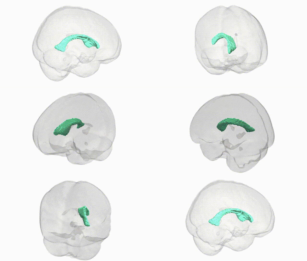
Full Quality Version: Download MP4
Wali Sidiqyar*, Gaurav Rudravaram*, Elyssa M. McMaster, Trent M. Schwartz, Adam M. Saunders, Kurt G. Schilling, Bennett A. Landman "Introducing SPINS: A Shared Public Visualization Library of Neuroanatomical Structures." Medical Imaging with Deep Learning- short paper
This resource is licensed under CC0 1.0 Universal (Public Domain).