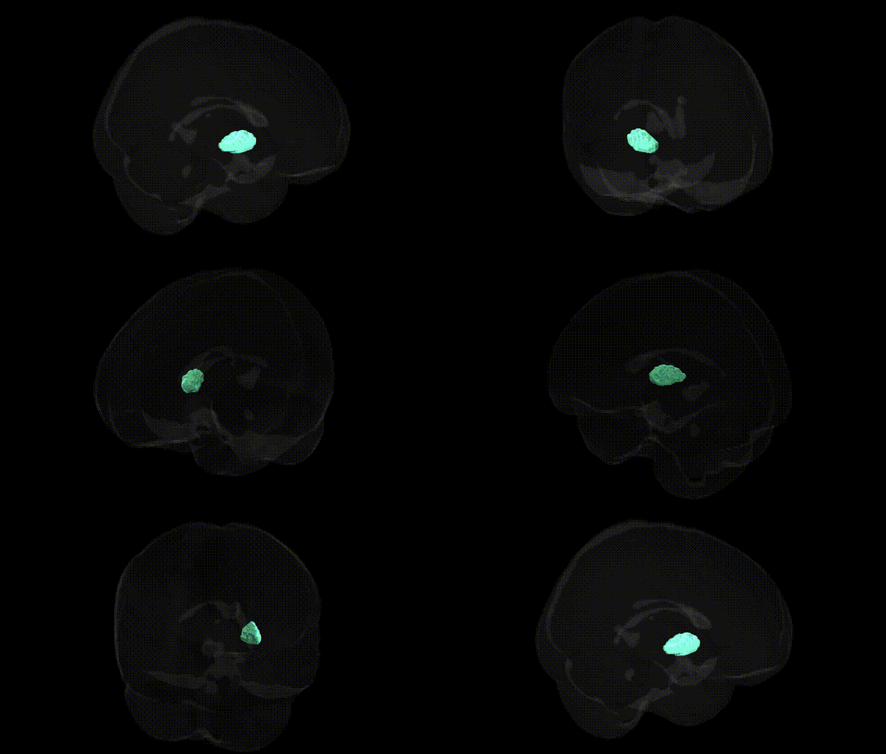
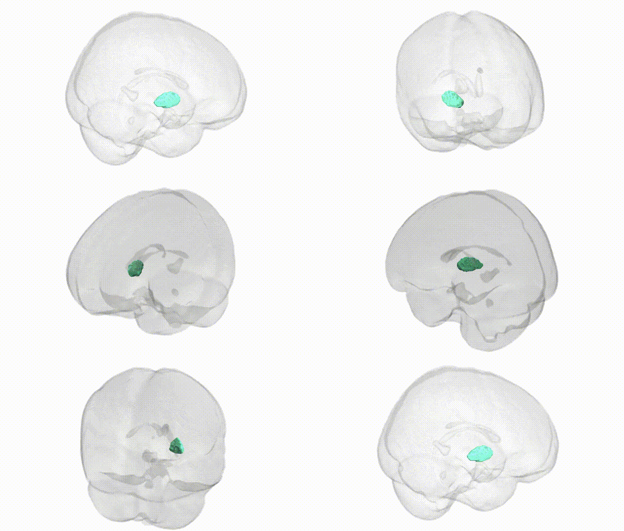
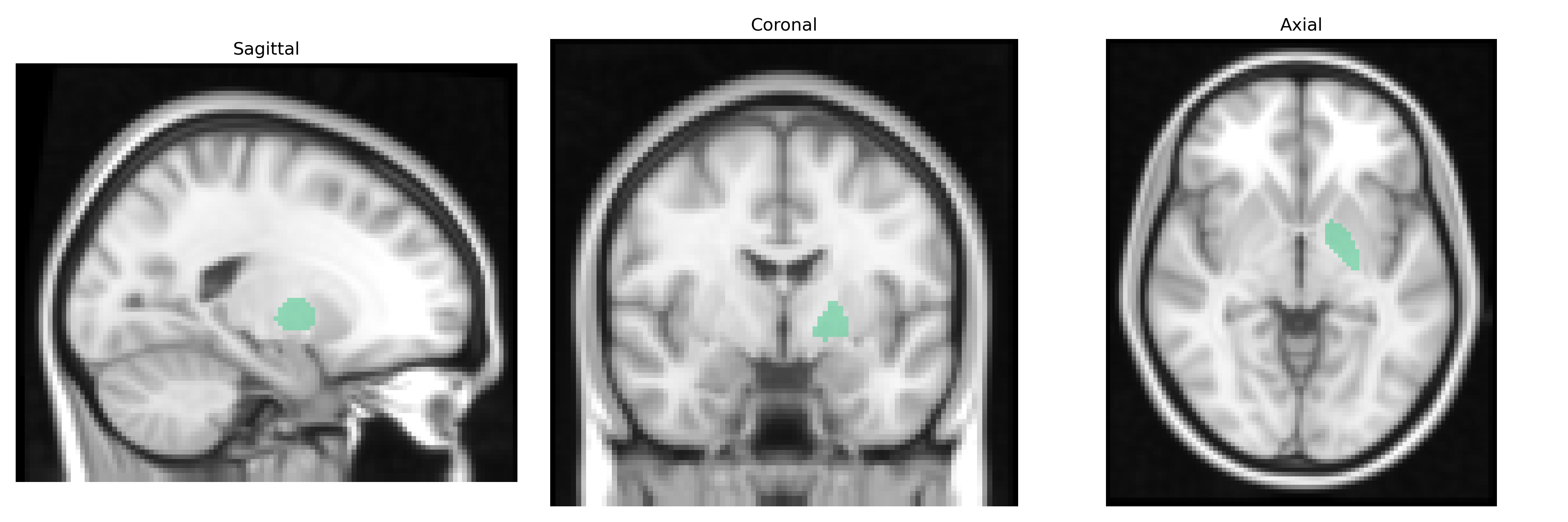
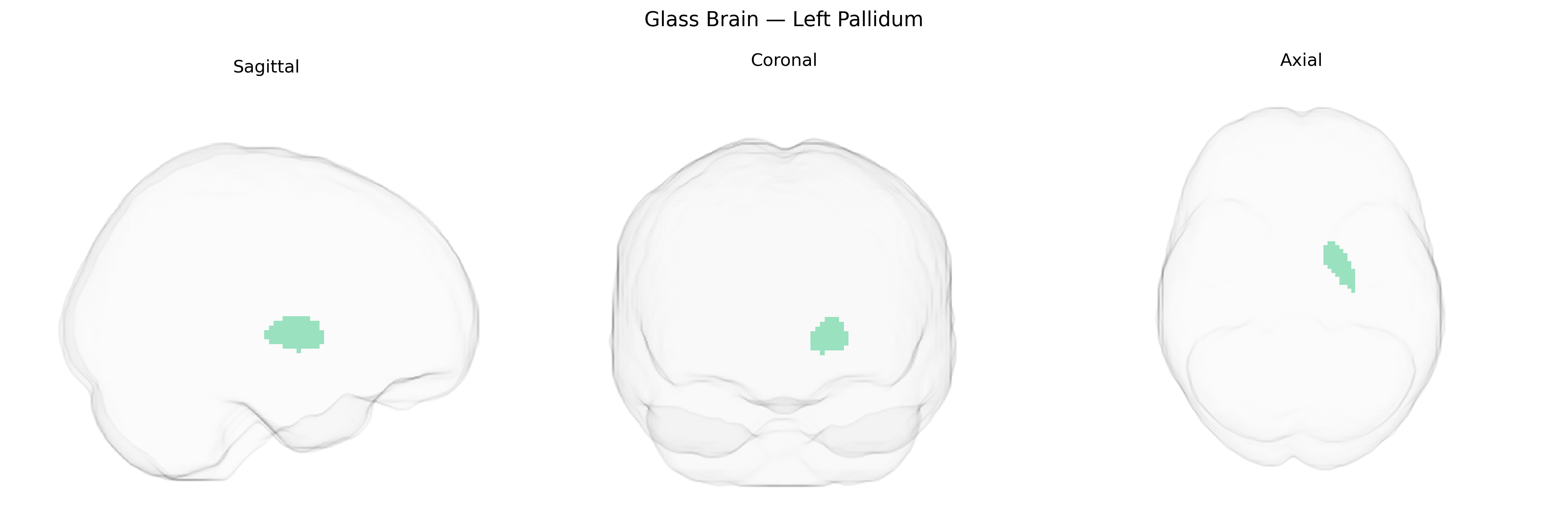
The bilateral Left Pallidum, as defined in the Harvard-Oxford subcortical maxprob thr25 2mm atlas, refers to the left-sided portion of the globus pallidus, a major component of the basal ganglia involved in the regulation of voluntary movement, muscle tone, and motor learning. This structure is subdivided into internal (GPi) and external (GPe) segments, which form critical nodes in cortico-striato-pallido-thalamo-cortical loops, integrating input from the striatum and modulating thalamic output to frontal cortical regions. Neurons in the pallidum are predominantly GABAergic, providing tonic inhibitory control that shapes the timing and selection of motor programs and also contributes to cognitive and affective processes. Pathophysiological alterations in the pallidum are implicated in movement disorders such as Parkinson’s disease and dystonia, making it a key target for interventions including deep brain stimulation. There is no direct Wikipedia article specifically for “Left Pallidum”; a closely related structure is the Globus pallidus.
The bilateral left pallidum, part of the basal ganglia, shows robust heritability and has been linked in large-scale imaging genetics and GWAS studies to variants in genes involved in neurodevelopment, synaptic function, and dopaminergic signaling, including loci near or within genes such as DCC, BDNF, and several glutamatergic and GABAergic pathway genes that influence subcortical volumes. GWAS of pallidum volume (e.g., ENIGMA, UK Biobank) have identified multiple common variants associated with its structural size, some of which overlap with risk loci for neuropsychiatric disorders such as schizophrenia, bipolar disorder, major depressive disorder, and obsessive-compulsive disorder, consistent with the pallidum’s role in cortico-striato-thalamo-cortical circuits. Genetic variants influencing pallidal structure and function have also been implicated in Parkinson’s disease, dystonia, and other movement disorders, where genes affecting dopaminergic transmission (e.g., DRD2-related pathways) and neurodegeneration (e.g., GBA and LRRK2 in broader basal ganglia contexts) show convergent evidence with pallidal involvement. Additionally, polygenic risk scores for traits such as cognitive performance, risk-taking, and addiction-related behaviors exhibit associations with pallidal morphology and activation, suggesting that shared genetic architecture links the pallidum to reward, motivation, and executive function. Overall, genetic studies support the pallidum as a heritable, genetically modulated hub whose structure and function are influenced by variants overlapping with multiple psychiatric, neurological, and behavioral phenotypes, even though specific GWAS are rarely limited to the left pallidum as defined in the HarvardOxford subcortical atlas and instead treat it as part of broader bilateral pallidal or basal ganglia measures.
Overview generated by GPT-4o (2026).
Region ID: 7
Hemisphere: bilateral
Atlas: HarvardOxford sub maxprob thr25 2mm
Full Quality Version: Download MP4
Full Quality Version: Download MP4
Wali Sidiqyar*, Gaurav Rudravaram*, Elyssa M. McMaster, Trent M. Schwartz, Adam M. Saunders, Kurt G. Schilling, Bennett A. Landman "Introducing SPINS: A Shared Public Visualization Library of Neuroanatomical Structures." Medical Imaging with Deep Learning- short paper
This resource is licensed under CC0 1.0 Universal (Public Domain).