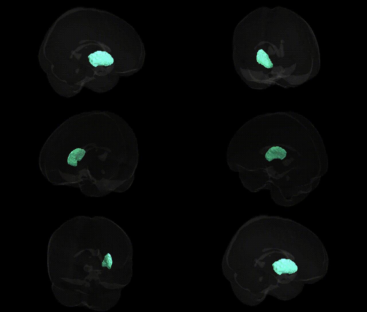
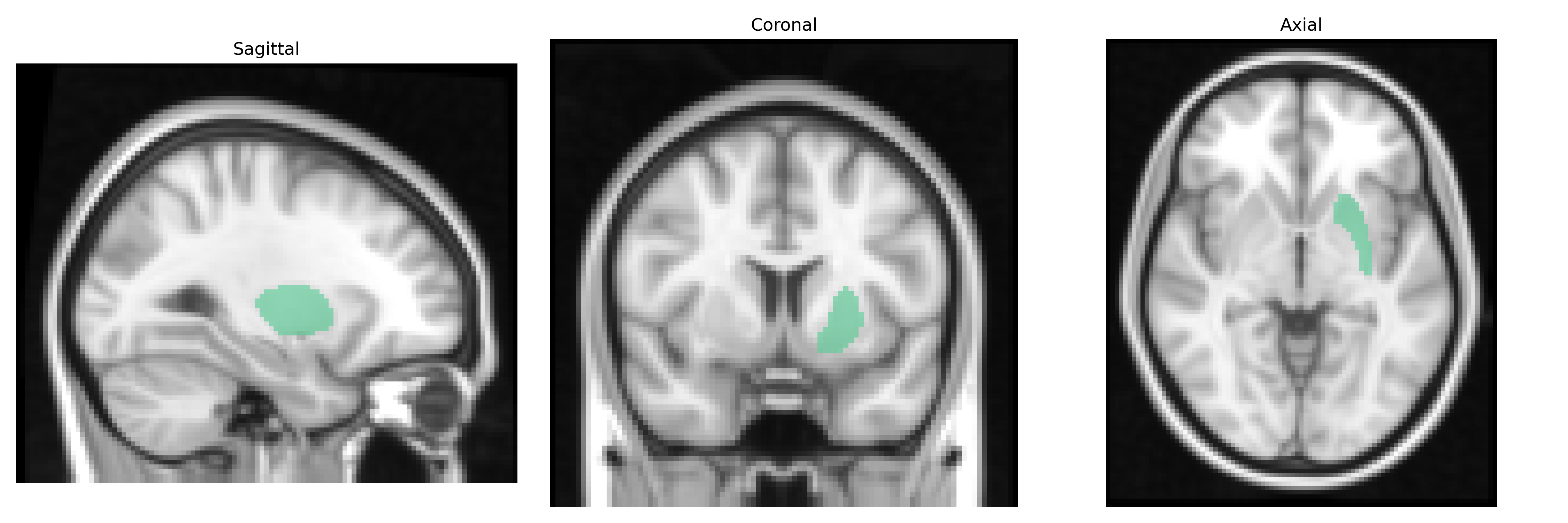
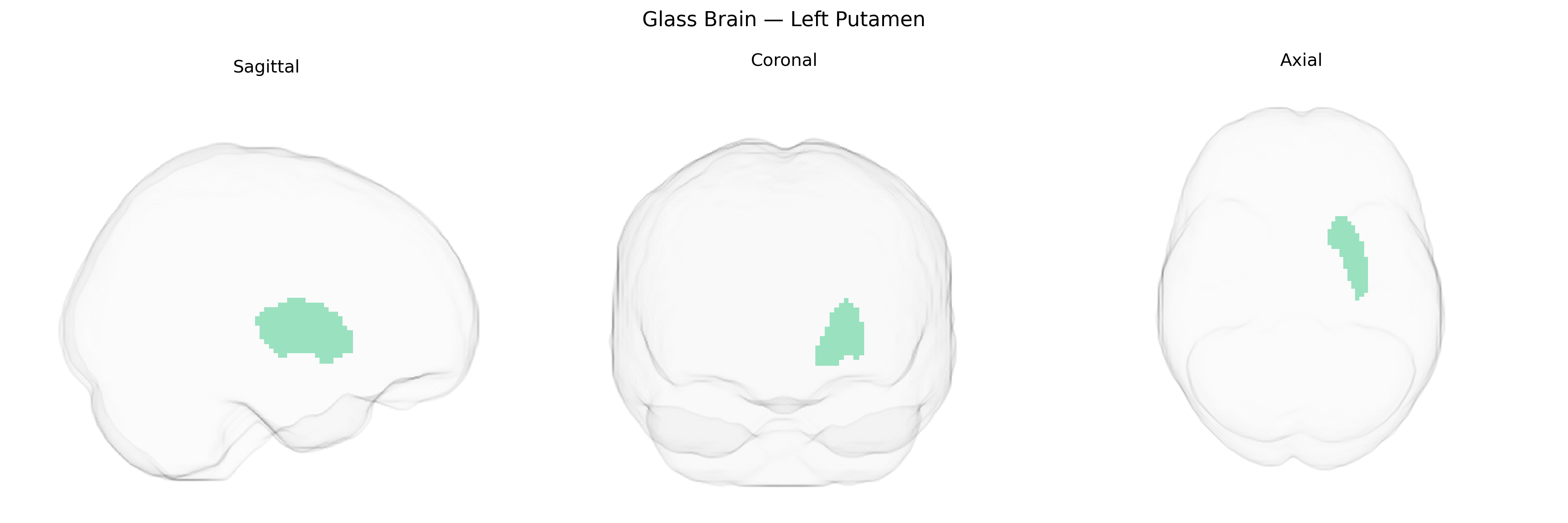
The bilateral Left Putamen, as defined in the Harvard-Oxford Subcortical (sub maxprob thr25 2mm) Atlas, is a paired deep gray matter structure within the dorsal striatum of the basal ganglia, located lateral to the globus pallidus and medial to the insular cortex. Composed predominantly of medium spiny GABAergic neurons receiving dense glutamatergic input from motor, premotor, and associative cortices, it serves as a major input nucleus of the basal ganglia circuitry. The putamen is critically involved in motor control, habit formation, procedural learning, and aspects of reward-related processing, integrating cortical signals and modulating thalamocortical loops that govern movement initiation, execution, and automaticity. Through its connectivity with the caudate nucleus, globus pallidus, substantia nigra, and subthalamic nucleus, the putamen contributes to both direct and indirect pathways that facilitate or inhibit motor output, and it is implicated in movement disorders such as Parkinson’s disease and dystonia, as well as in certain neuropsychiatric conditions. Putamen
The left putamen, as defined in the Harvard-Oxford subcortical atlas, has been implicated in numerous genetic studies, particularly GWAS of neuropsychiatric and neurodegenerative traits, reflecting its role in motor, reward, and habit circuitry. Variants in genes affecting striatal development, synaptic function, and dopaminergic signaling (e.g., DRD2, COMT, BDNF, and multiple loci identified in large imaging-genetics consortia such as ENIGMA and UK Biobank) have been associated with putamen volume and microstructure, with several loci showing bilateral effects that include the left putamen. GWAS have linked left (and bilateral) putamen morphology to schizophrenia, bipolar disorder, major depressive disorder, obsessive–compulsive disorder, attention-deficit/hyperactivity disorder, and autism spectrum disorder, as well as to substance use traits (including alcohol and tobacco use) and personality dimensions related to impulsivity and neuroticism, often via polygenic risk scores that correlate with putaminal structure and function. In movement disorders, genes central to dopaminergic and basal ganglia pathways, such as those associated with Parkinson’s disease (e.g., SNCA, LRRK2 and other PD risk loci) and monogenic dystonias, show downstream structural and functional consequences in the putamen. Additionally, imaging GWAS have identified common variants influencing left putamen volume that overlap with loci for cognitive performance, educational attainment, and general brain size, highlighting shared genetic architecture between striatal anatomy and broader cognitive and psychiatric phenotypes.
Overview generated by GPT-4o (2026).
Region ID: 6
Hemisphere: bilateral
Atlas: HarvardOxford sub maxprob thr25 2mm
Full Quality Version: Download MP4
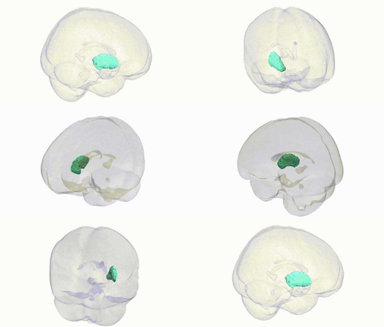
Full Quality Version: Download MP4
Wali Sidiqyar*, Gaurav Rudravaram*, Elyssa M. McMaster, Trent M. Schwartz, Adam M. Saunders, Kurt G. Schilling, Bennett A. Landman "Introducing SPINS: A Shared Public Visualization Library of Neuroanatomical Structures." Medical Imaging with Deep Learning- short paper
This resource is licensed under CC0 1.0 Universal (Public Domain).