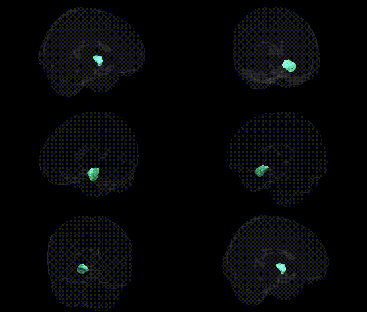
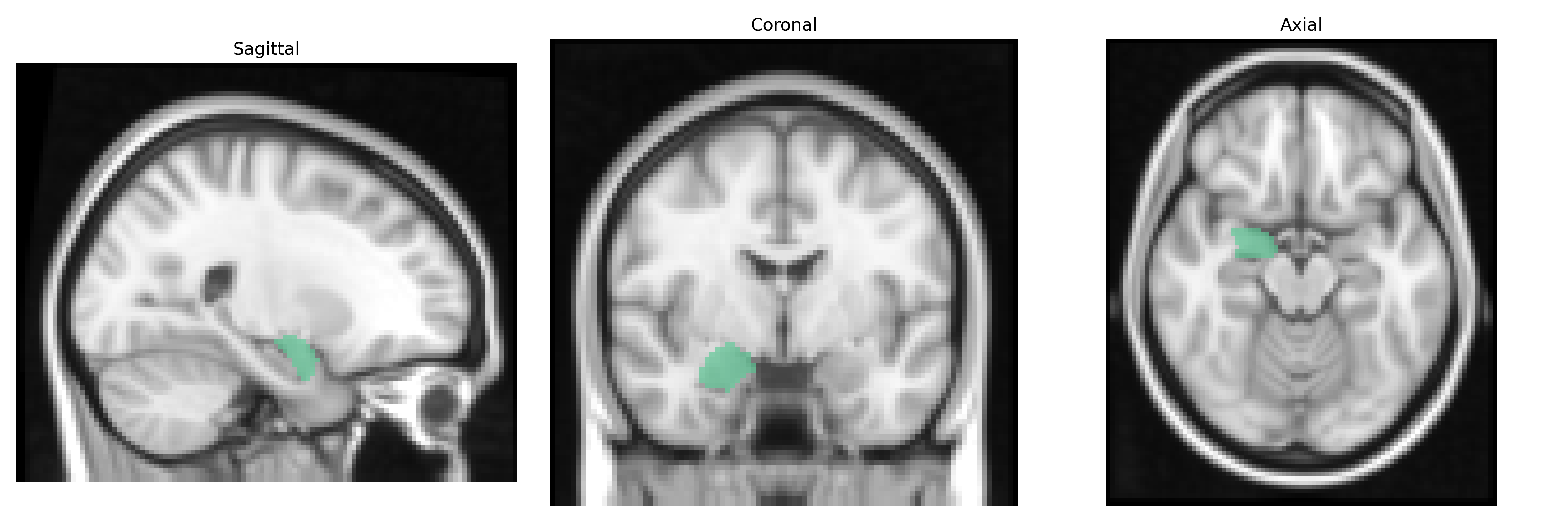
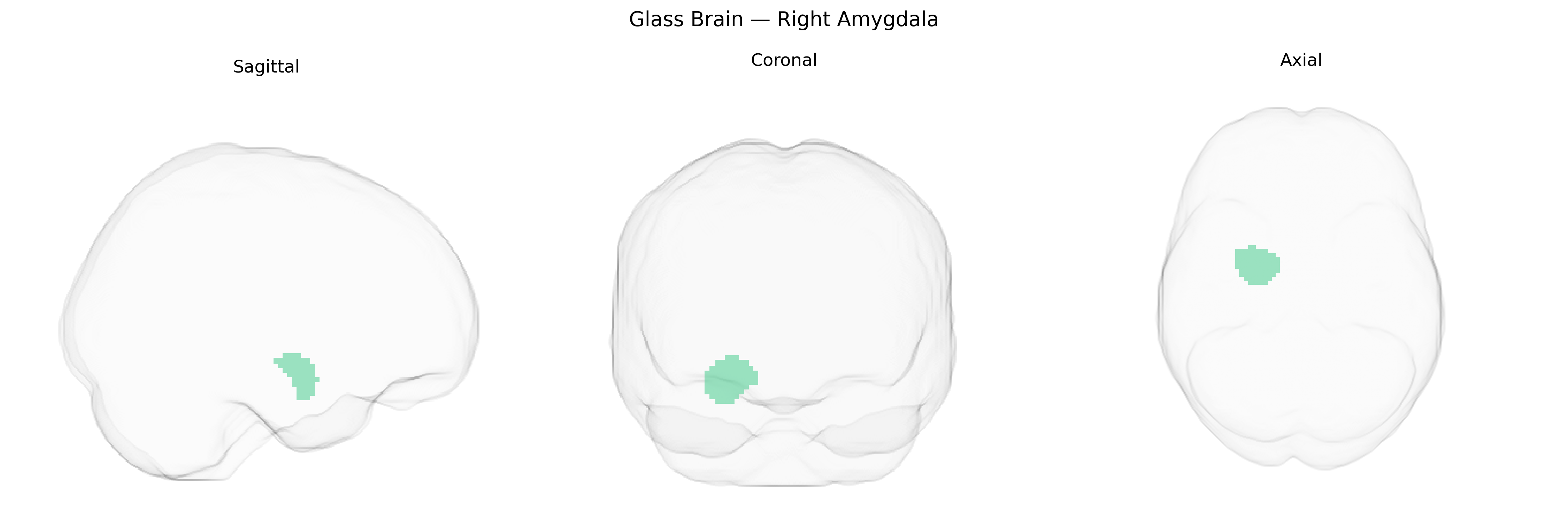
The bilateral Right Amygdala, as defined in the Harvard-Oxford subcortical maximum probability (25%, 2 mm) atlas, refers to the probability-based delineation of amygdaloid gray matter within the right medial temporal lobe, mirrored across hemispheres for bilateral analyses. This region encompasses the main amygdaloid nuclei complex, including basolateral, centromedial, and cortical subdivisions, which are interconnected with the hippocampus, prefrontal cortex, hypothalamus, and brainstem. Functionally, the amygdala is critically involved in emotional processing, especially fear and threat detection, associative emotional learning, modulation of autonomic and endocrine responses, and influence over memory consolidation for emotionally salient events. In neuroimaging, this atlas label is commonly used to quantify activation, volume, or connectivity in studies of affective processing, anxiety, mood disorders, and social cognition. Amygdala
The bilateral right amygdala, as defined in the Harvard-Oxford subcortical atlas, has been implicated in numerous genetic studies, particularly GWAS of brain structure and psychiatric traits. Variants near MEF2C, SLC39A8, and HMGA2, as well as loci in the 8q24 and 12q23 regions, have been associated with amygdala volume or morphology, with several large neuroimaging-genetics consortia (e.g., ENIGMA) reporting polygenic influences on right amygdala size that partially overlap with genetic architectures for major depressive disorder, schizophrenia, bipolar disorder, and anxiety-related traits. Risk alleles in genes involved in synaptic plasticity and stress response, including BDNF (e.g., Val66Met), FKBP5, CRHR1, and serotonin-related genes (such as SLC6A4/5-HTTLPR), have been linked to altered amygdala reactivity and functional connectivity, often with lateralized effects favoring right amygdala hyperactivation in response to emotional or threatening stimuli. GWAS of neuroticism, social anxiety, post-traumatic stress disorder, and aggression-related traits show enriched associations in networks encompassing the amygdala, and polygenic risk scores for these disorders correlate with altered right amygdala volume and activity. In addition, Alzheimer’s disease and other neurodegenerative GWAS highlight loci (e.g., APOE, CLU, BIN1) that are associated with amygdala atrophy patterns, with some evidence that right-sided amygdala changes are particularly sensitive to genetic risk burden, reflecting the broader role of this region as a genetically modulated hub for emotional processing, threat detection, and memory-related psychopathology.
Overview generated by GPT-4o (2026).
Region ID: 20
Hemisphere: bilateral
Atlas: HarvardOxford sub maxprob thr25 2mm
Full Quality Version: Download MP4
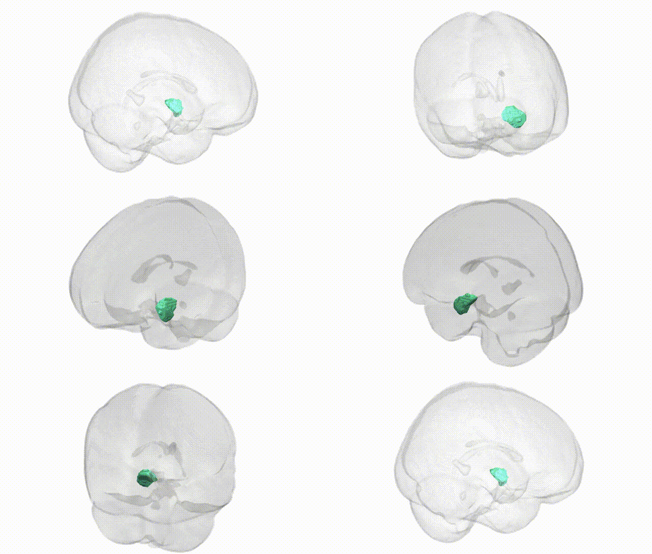
Full Quality Version: Download MP4
Wali Sidiqyar*, Gaurav Rudravaram*, Elyssa M. McMaster, Trent M. Schwartz, Adam M. Saunders, Kurt G. Schilling, Bennett A. Landman "Introducing SPINS: A Shared Public Visualization Library of Neuroanatomical Structures." Medical Imaging with Deep Learning- short paper
This resource is licensed under CC0 1.0 Universal (Public Domain).