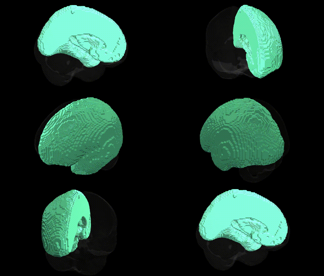
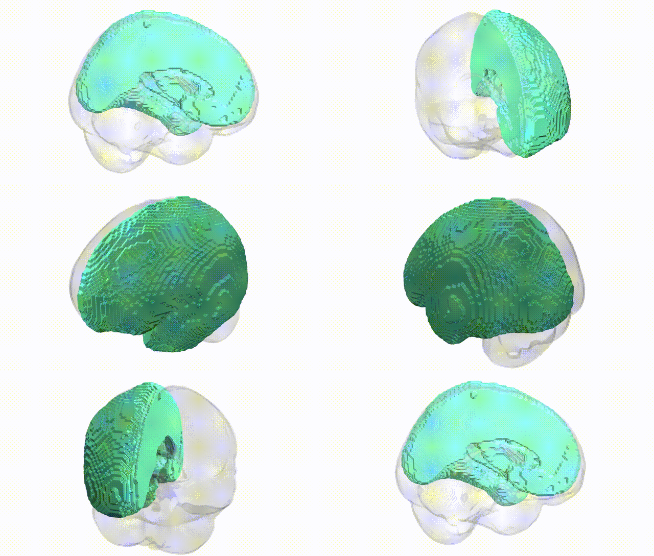
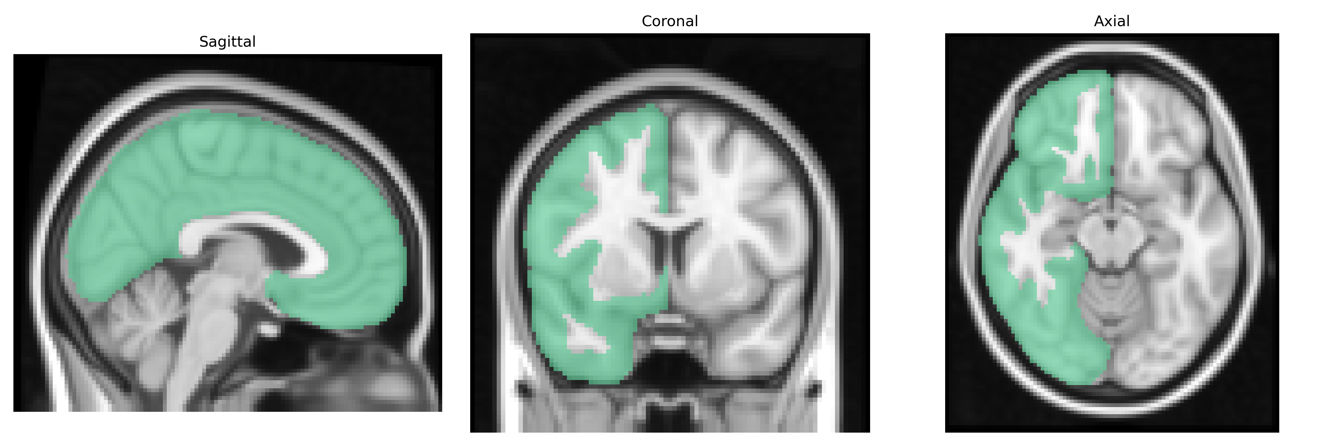
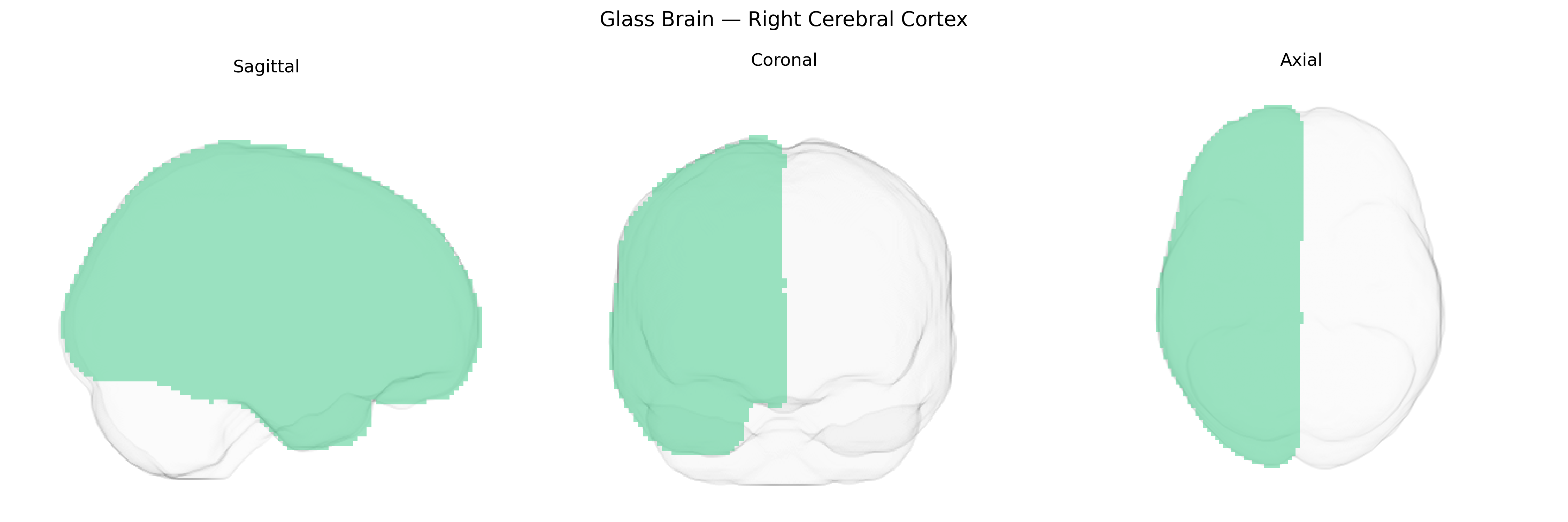
The bilateral right cerebral cortex, as defined in the Harvard-Oxford subcortical maximum probability atlas (thr25, 2 mm), encompasses the entire mantle of gray matter covering the right cerebral hemisphere, including frontal, parietal, temporal, and occipital cortices, as well as insular regions. This structure consists primarily of neuronal cell bodies organized into six-layered neocortex, with regional cytoarchitectonic and connectivity specializations that support higher-order cognitive processes (e.g., attention, executive function, language prosody), sensory perception (somatosensory, visual, auditory), motor planning and control, and integration of multimodal information. Functionally, the right cerebral cortex is often associated with visuospatial processing, holistic perception, aspects of social cognition, and nonverbal communication, while maintaining extensive reciprocal connections with subcortical structures (thalamus, basal ganglia, limbic system) and the left hemisphere via the corpus callosum. There is no direct Wikipedia article for “Right Cerebral Cortex”; a closely related general entry is Cerebral cortex.
Genetic associations with the bilateral right cerebral cortex, as broadly defined in the Harvard–Oxford subcortical/maxprob atlas, derive mainly from large-scale GWAS of cortical volume, thickness, and surface area that treat the right hemisphere either globally or by lobar/subregional parcellations. Variants in genes regulating neurodevelopment and synaptic function—such as HMGA2, MIR588, LINC01122, and loci near IGF1 and WNT signaling components—have been linked to global or hemisphere-specific cortical metrics, often with asymmetric effects favoring right-hemispheric associations. Many schizophrenia, bipolar disorder, and major depression risk loci (e.g., in CACNA1C, ZNF804A, GRIN2A) show downstream effects on right frontal and temporal cortical thickness and surface area, while autism spectrum disorder–associated genes (e.g., CHD8, SHANK3, NRXN1) have been implicated in altered right cortical morphology and connectivity, particularly in temporal and parietal association areas. GWAS of cognitive traits and educational attainment (e.g., involving variants near KIAA0319, FOXP2-related networks, and polygenic scores for intelligence) reveal correlations with right cortical volume and surface area, especially in right frontal and parietotemporal regions involved in attention, visuospatial processing, and higher-order cognition. Additionally, polygenic risk for neurodegenerative diseases such as Alzheimer’s (APOE, BIN1, CLU) associates with right cortical thinning in temporal and parietal cortex, and migraine-, epilepsy-, and ADHD-related loci (e.g., near TRPM8, SCN1A, DRD4) have been tied to structural and functional alterations in right cortical networks, underscoring a broad genetic influence on this hemisphere’s structure and disease vulnerability even when not explicitly labeled as “right cerebral cortex” in atlas-based analyses.
Overview generated by GPT-4o (2026).
Region ID: 13
Hemisphere: bilateral
Atlas: HarvardOxford sub maxprob thr25 2mm
Full Quality Version: Download MP4
Full Quality Version: Download MP4
Wali Sidiqyar*, Gaurav Rudravaram*, Elyssa M. McMaster, Trent M. Schwartz, Adam M. Saunders, Kurt G. Schilling, Bennett A. Landman "Introducing SPINS: A Shared Public Visualization Library of Neuroanatomical Structures." Medical Imaging with Deep Learning- short paper
This resource is licensed under CC0 1.0 Universal (Public Domain).