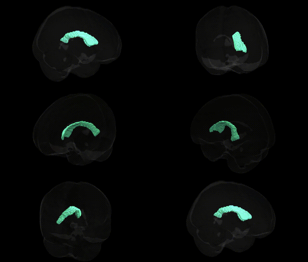
The bilateral right lateral ventricle, as defined in the Harvard-Oxford subcortical maximum probability (25% threshold, 2 mm) atlas, refers to the cerebrospinal fluid (CSF)-filled cavity within the right cerebral hemisphere that forms part of the ventricular system. It comprises the anterior (frontal) horn, body, atrium (trigone), and posterior (occipital) and inferior (temporal) horns, each following the corresponding lobes of the right hemisphere. The lateral ventricle is lined by ependymal cells and bordered by critical structures including the caudate nucleus, corpus callosum, thalamus, and hippocampus, making it a key neuroanatomical landmark for structural and functional imaging. It participates in CSF production and circulation via the choroid plexus and communicates with the third ventricle through the interventricular foramen (of Monro). There is no direct Wikipedia article for “Right Lateral Ventricle”; a related and encompassing structure is the Lateral ventricle.
Genetic associations related to the bilateral Right Lateral Ventricle—operationalized in many neuroimaging genetics studies as lateral ventricular volume rather than atlas-specific parcellations—have been identified primarily through large-scale GWAS of brain structure. Variants in genes such as APOE (particularly in the context of Alzheimer’s disease), MAPT, and GRIN2B, along with loci near MIR9-2, have been repeatedly linked to ventricular enlargement, which is often interpreted as a marker of global or regional brain atrophy rather than a direct functional substrate. GWAS from consortia like ENIGMA and UK Biobank have found polygenic influences on lateral ventricular size that overlap with genetic architectures for neurodegenerative disorders, schizophrenia, bipolar disorder, and cognitive traits, suggesting that risk alleles for these conditions tend to be associated with increased ventricular volume, especially in the presence of cortical and subcortical tissue loss. Additional loci implicated in general brain development and intracranial volume (e.g., HMGA2, IGF1-related pathways, and other neurodevelopmental genes) also show associations with ventricular measures, indicating that genetic variants influencing neurodevelopment and neurodegeneration contribute to inter-individual differences in the right lateral ventricle as defined in structural MRI atlases such as the Harvard–Oxford subcortical atlas.
Overview generated by GPT-4o (2026).
Region ID: 14
Hemisphere: bilateral
Atlas: HarvardOxford sub maxprob thr25 2mm
Full Quality Version: Download MP4
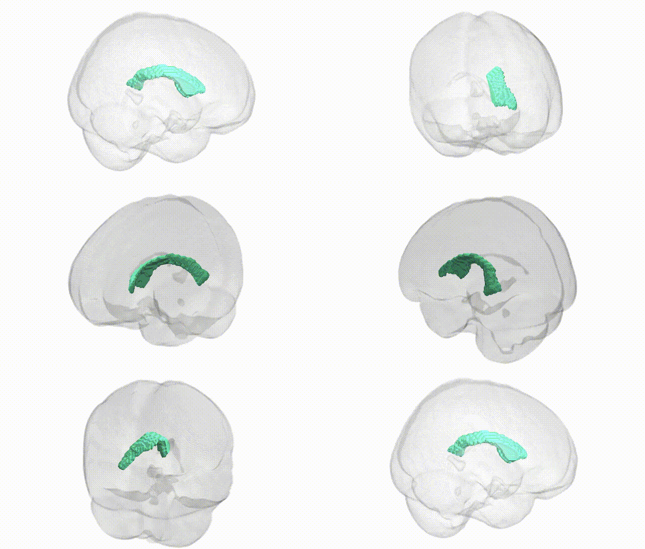
Full Quality Version: Download MP4
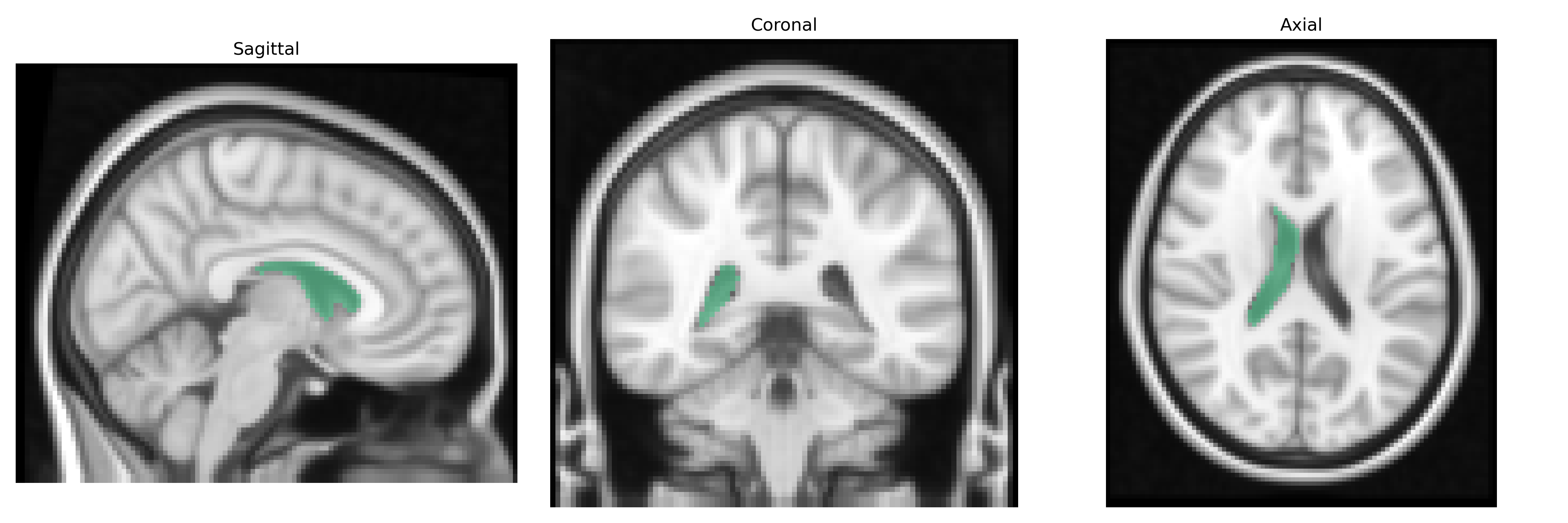
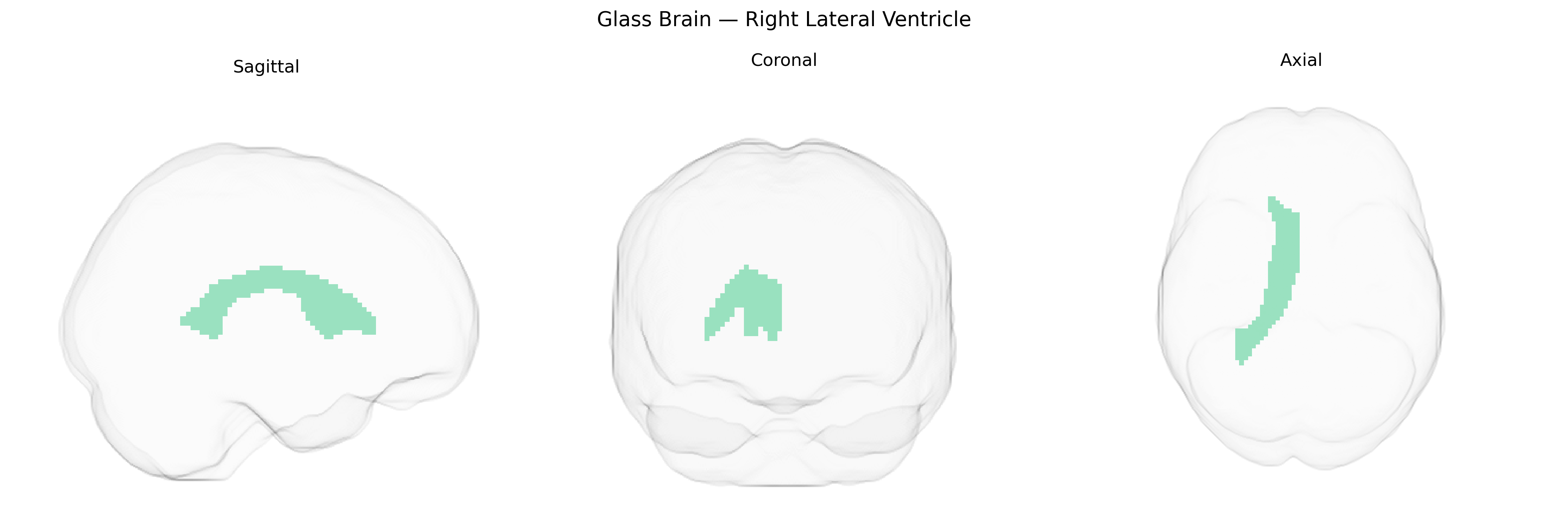
Wali Sidiqyar*, Gaurav Rudravaram*, Elyssa M. McMaster, Trent M. Schwartz, Adam M. Saunders, Kurt G. Schilling, Bennett A. Landman "Introducing SPINS: A Shared Public Visualization Library of Neuroanatomical Structures." Medical Imaging with Deep Learning- short paper
This resource is licensed under CC0 1.0 Universal (Public Domain).