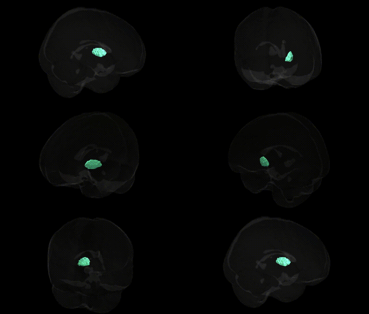
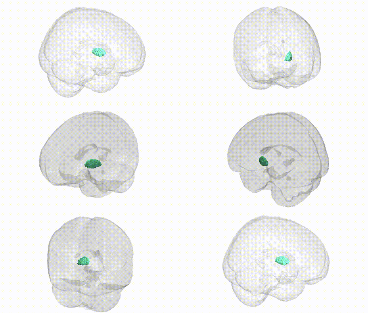
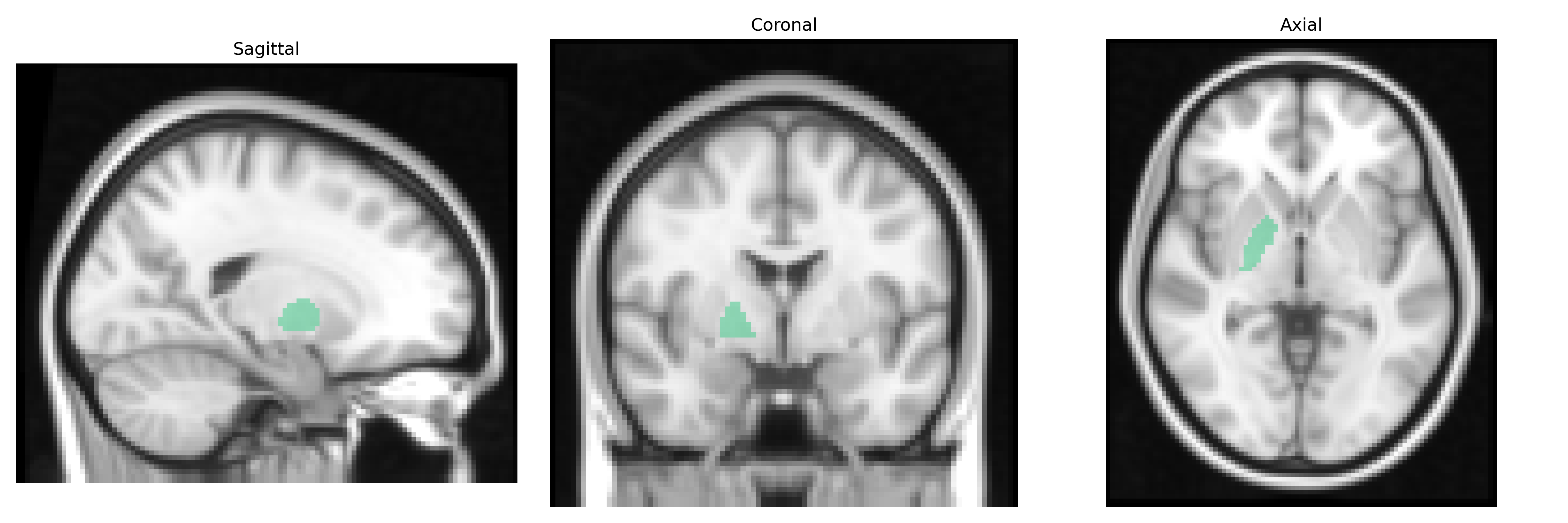
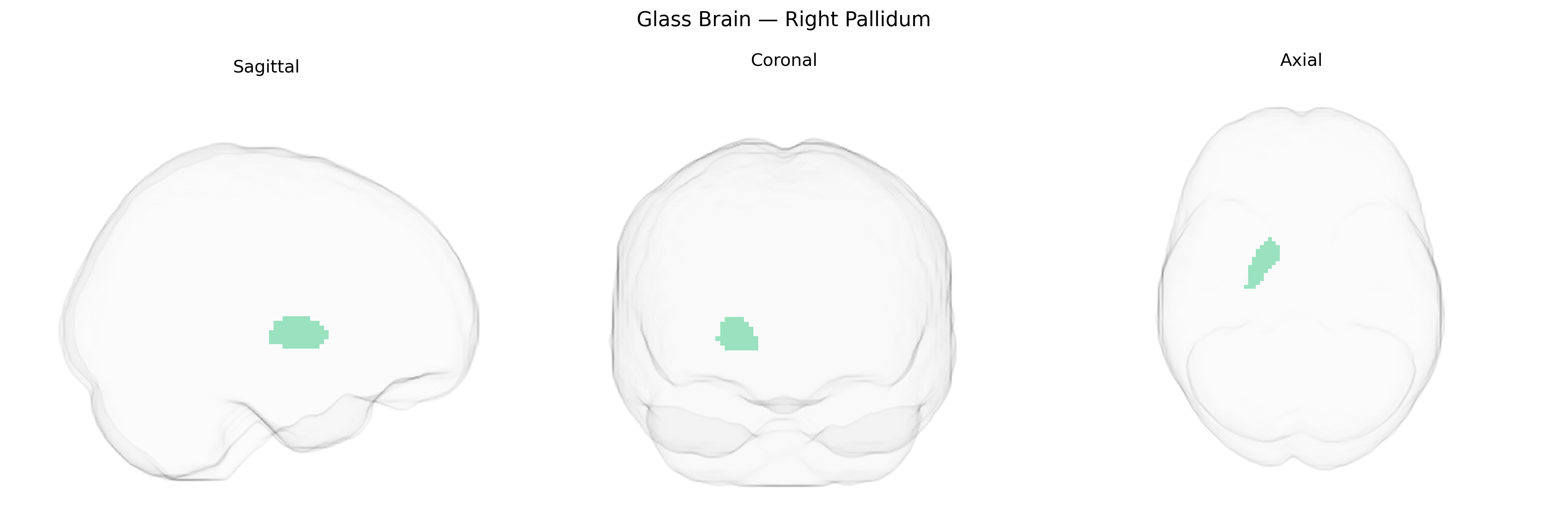
The bilateral right pallidum, as defined in the Harvard-Oxford Subcortical Structural Atlas (sub maxprob thr25, 2 mm), corresponds primarily to the right globus pallidus, a major component of the basal ganglia involved in the regulation of voluntary movement and motor tone. Structurally, the pallidum is divided into internal (medial) and external (lateral) segments, which receive inhibitory input mainly from the striatum and project via GABAergic neurons to thalamic nuclei and brainstem motor centers, thereby modulating cortical motor circuits. Functionally, this region is crucial for the refinement of movement, suppression of unwanted motor activity, and the integration of motor, cognitive, and limbic information, with dysfunction or lesion commonly associated with movement disorders such as Parkinson’s disease and dystonia. There is no direct Wikipedia article for “Right Pallidum”; a related structure is the Globus pallidus.
The bilateral right pallidum, part of the basal ganglia and mapped in the Harvard-Oxford subcortical atlas, has been implicated in multiple genetic studies, particularly GWAS of subcortical brain volumes and neuropsychiatric traits. Large-scale imaging genetics work (e.g., ENIGMA, UK Biobank) has identified SNPs in and around genes such as KTN1, DCC, DRD2, and variants near glutamatergic and GABAergic signaling genes that are associated with pallidal volume and morphology, often shared across left and right hemispheres. Pallidum-related genetic architecture overlaps with loci implicated in Parkinson’s disease, dystonia, schizophrenia, major depression, attention-deficit/hyperactivity disorder, and obsessive-compulsive disorder, where polygenic risk scores and GWAS hits in dopaminergic, synaptic, and neuronal development pathways track with functional abnormalities in basal ganglia circuits. Additionally, genetic variants influencing pallidum structure and activity have been linked to traits such as motor skill learning, reward sensitivity, cognitive performance, and addiction vulnerability, reflecting the region’s role in motor control and motivational processing. While many GWAS treat pallidum bilaterally or do not resolve hemisphere-specific effects, available data suggest that the genetic determinants of right pallidum volume and connectivity substantially overlap with those of the pallidum more generally and converge on pathways involved in neurodevelopment, synaptic plasticity, and dopaminergic modulation.
Overview generated by GPT-4o (2026).
Region ID: 18
Hemisphere: bilateral
Atlas: HarvardOxford sub maxprob thr25 2mm
Full Quality Version: Download MP4
Full Quality Version: Download MP4
Wali Sidiqyar*, Gaurav Rudravaram*, Elyssa M. McMaster, Trent M. Schwartz, Adam M. Saunders, Kurt G. Schilling, Bennett A. Landman "Introducing SPINS: A Shared Public Visualization Library of Neuroanatomical Structures." Medical Imaging with Deep Learning- short paper
This resource is licensed under CC0 1.0 Universal (Public Domain).