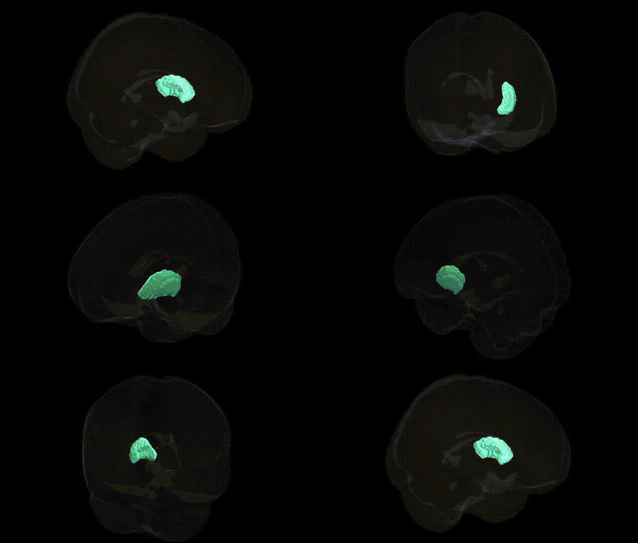
The bilateral Right Putamen, as defined in the Harvard-Oxford subcortical maxprob thr25 2mm atlas, is a paired deep gray matter structure within the dorsal striatum of the basal ganglia, located lateral to the globus pallidus and medial to the insular cortex. Composed predominantly of medium spiny GABAergic neurons, it receives dense glutamatergic input from motor, premotor, and associative cortices, as well as dopaminergic projections from the substantia nigra pars compacta. Functionally, it participates in cortico-basal ganglia-thalamo-cortical loops that modulate motor planning, execution, and habit formation, and it contributes to aspects of associative learning, reward processing, and procedural memory. Structural and functional alterations of the putamen are implicated in movement disorders such as Parkinson’s disease and Huntington’s disease, as well as in various neuropsychiatric conditions involving frontostriatal circuitry.
The bilateral right putamen, a core dorsal striatal structure in the Harvard-Oxford subcortical atlas, has been repeatedly implicated in genetic studies of motor control, addiction, and neuropsychiatric disorders. Large-scale GWAS of brain imaging phenotypes (e.g., ENIGMA, UK Biobank) have identified loci near genes such as CAV2, BDNF, DRD2, GMNC, and HRK associated with putamen volume, reflecting its sensitivity to variants in neurodevelopmental, synaptic, and dopaminergic pathways. Genetic risk for Parkinson’s disease (e.g., SNCA, LRRK2, GBA variants) and Huntington’s disease (HTT CAG expansions) converges on striatal circuits including the putamen, consistent with its central role in motor dysfunction and degeneration. Schizophrenia and bipolar disorder polygenic risk scores, as well as specific loci such as CACNA1C and those in glutamatergic and GABAergic systems, have been associated with altered putamen structure and function, supporting a genetic contribution to cortico-striatal dysconnectivity. GWAS of substance use and dependence (notably alcohol, nicotine, and stimulant use) reveal overlap between addiction-risk loci and genes that modulate dopaminergic signaling in the putamen, paralleling findings from imaging genetics where allelic variation in DRD2/ANKK1, OPRM1, and related receptors influences reward-related activation of this region. Additionally, polygenic influences on personality traits (e.g., neuroticism, impulsivity) and cognitive functions show associations with putamen morphology and connectivity, suggesting that common genetic variation affecting corticostriatal development and neurotransmission partly shapes individual differences in behavior and vulnerability to psychiatric and neurological disorders.
Overview generated by GPT-4o (2026).
Region ID: 17
Hemisphere: bilateral
Atlas: HarvardOxford sub maxprob thr25 2mm
Full Quality Version: Download MP4
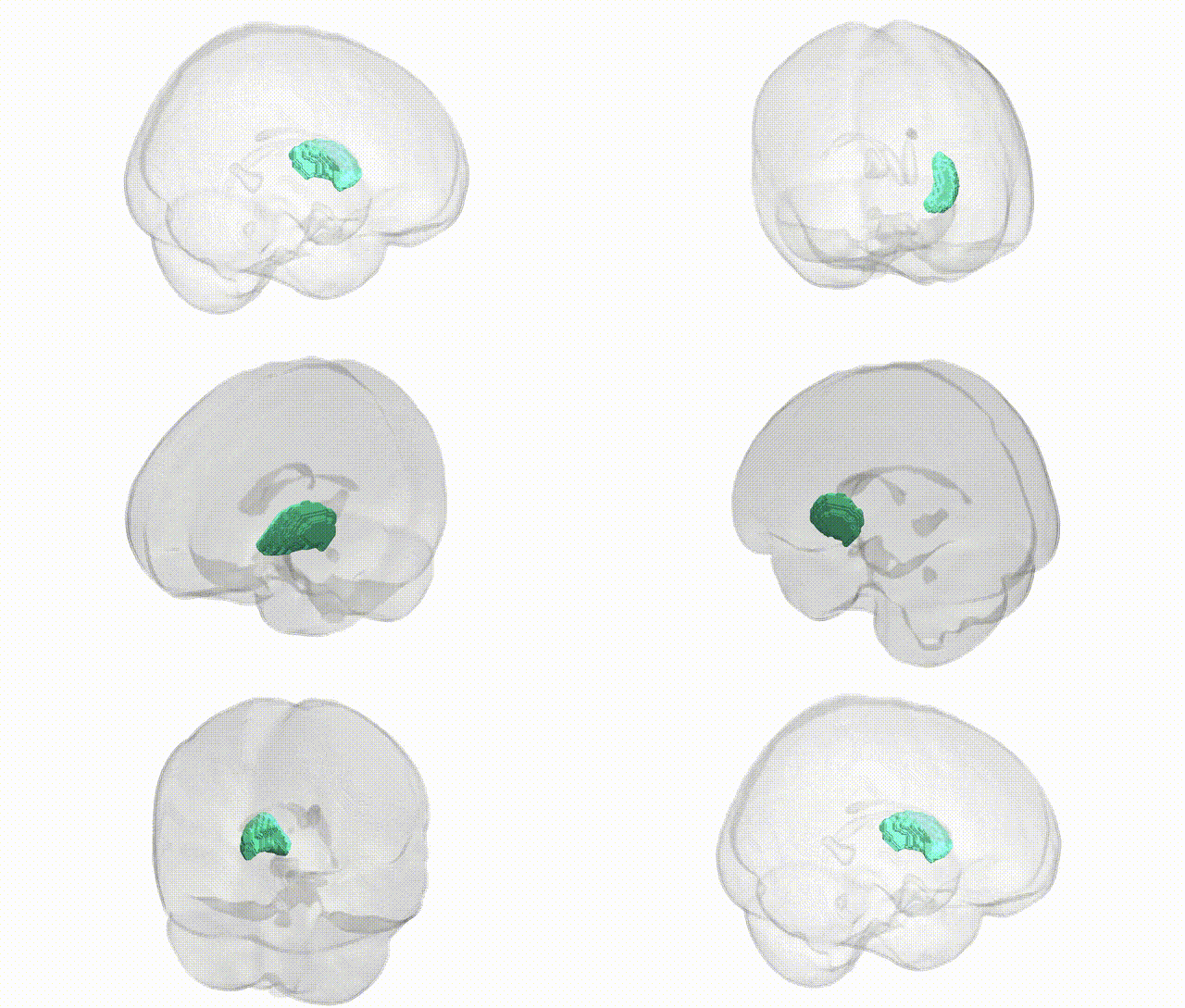
Full Quality Version: Download MP4
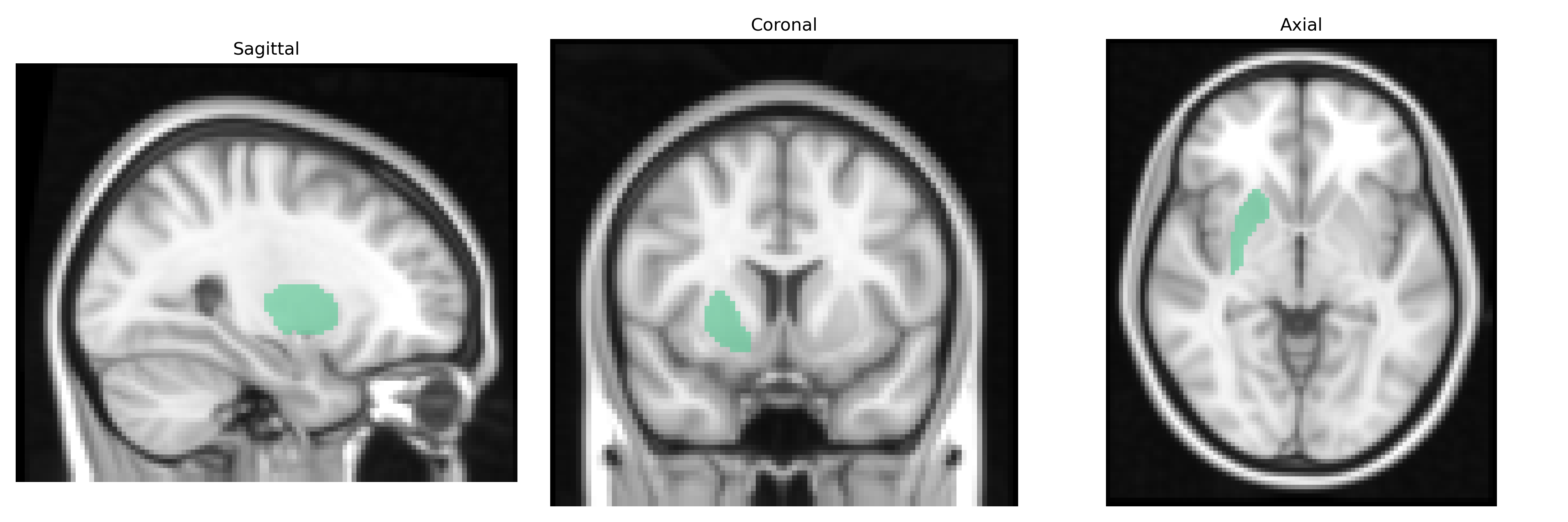
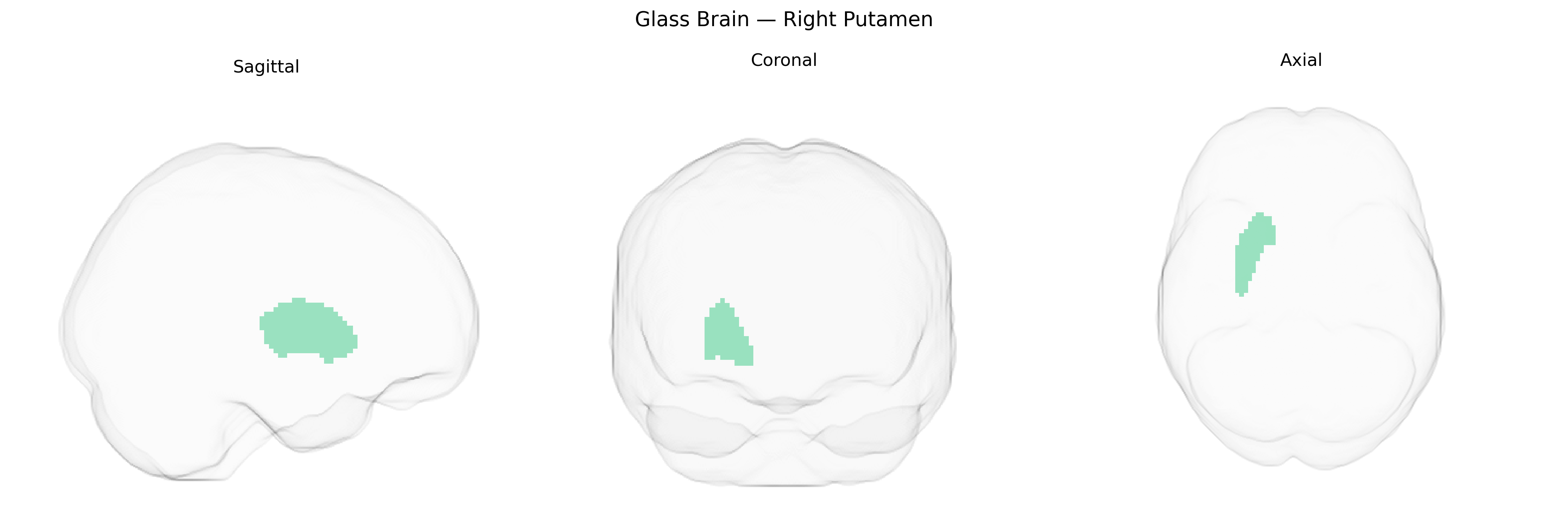
Wali Sidiqyar*, Gaurav Rudravaram*, Elyssa M. McMaster, Trent M. Schwartz, Adam M. Saunders, Kurt G. Schilling, Bennett A. Landman "Introducing SPINS: A Shared Public Visualization Library of Neuroanatomical Structures." Medical Imaging with Deep Learning- short paper
This resource is licensed under CC0 1.0 Universal (Public Domain).