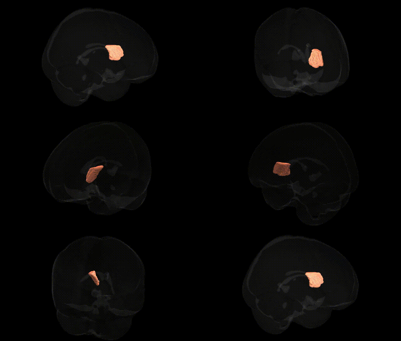
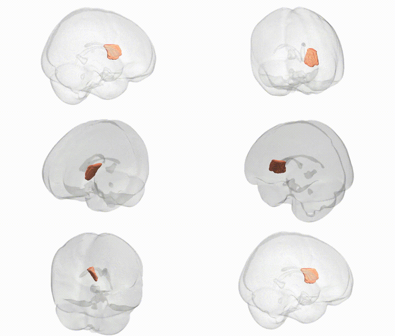
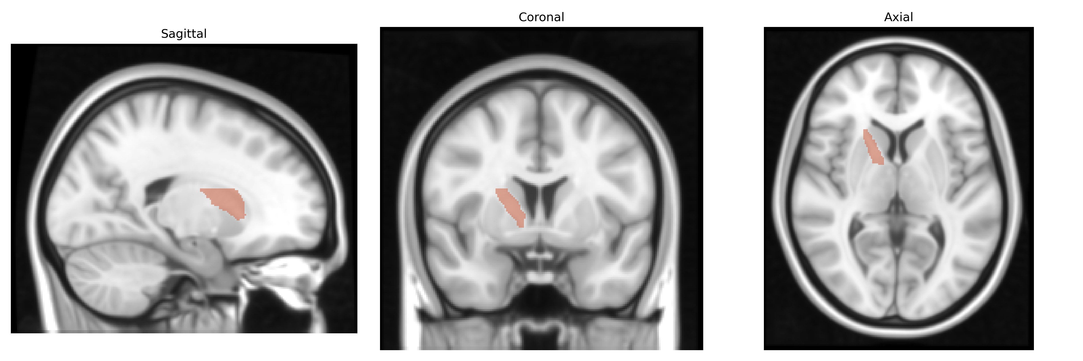
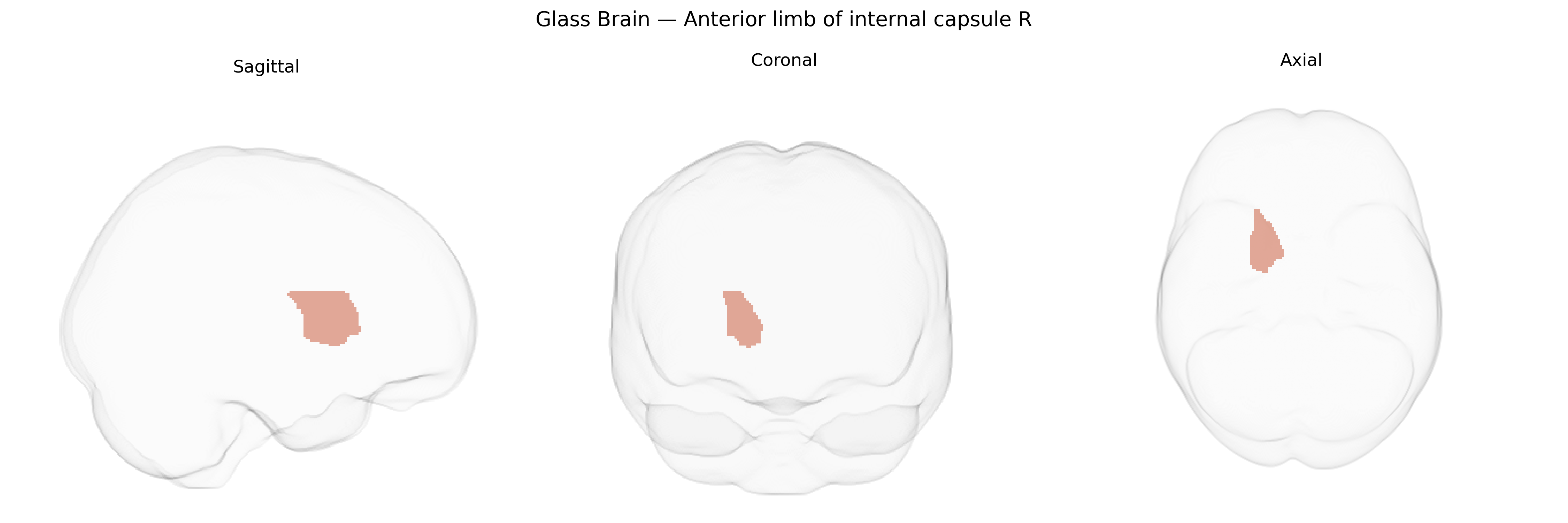
The bilateral Anterior limb of internal capsule R, as defined in the JHU ICBM labels 1 mm Atlas, corresponds to the right-sided anterior portion of the internal capsule, a major white matter tract situated between the head of the caudate nucleus medially and the lentiform nucleus laterally. It contains frontopontine fibers, thalamocortical projections (particularly from mediodorsal and anterior thalamic nuclei to prefrontal and limbic cortices), and reciprocal cortico-thalamic connections, thereby playing key roles in executive function, motivation, and affective processing. This region is crucial for large-scale network communication between frontal association areas and subcortical structures, and its microstructural integrity is frequently assessed in diffusion MRI studies of neuropsychiatric, neurodegenerative, and cerebrovascular disorders. There is no direct Wikipedia article for this specific labeled region; a closely related structure is the Internal capsule.
The bilateral anterior limb of the internal capsule (ALIC), as labeled in the JHU ICBM 1 mm Atlas, has been implicated in multiple genetic and imaging-genetic studies, though typically as part of broader white matter or fronto-striatal circuitry rather than as a uniquely isolated tract. Genome-wide association studies (GWAS) of diffusion tensor imaging metrics (e.g., fractional anisotropy, mean diffusivity) have identified significant heritability and common variant influences on ALIC microstructure, with loci involving axon guidance, myelination, and neurodevelopmental pathways (such as genes in the netrin, semaphorin, and neuregulin signaling families) contributing to inter-individual variability. Polygenic risk scores for schizophrenia, bipolar disorder, and major depressive disorder have been associated with altered ALIC integrity, consistent with the role of the tract in cortico-striatal-thalamic loops linked to mood, motivation, and cognitive control. Variants in genes related to myelin (e.g., MAG, MBP-related networks), neuroinflammation (e.g., HLA region), and synaptic plasticity have been reported in imaging genetics work to modulate ALIC structure or connectivity, aligning with findings of white matter abnormalities in psychiatric and neurodevelopmental disorders, including schizophrenia, obsessive-compulsive disorder, and attention-deficit/hyperactivity disorder. In addition, neurodegenerative and vascular GWAS (involving loci such as APOE and genes influencing small-vessel disease) have been connected to microstructural changes in internal capsule segments, including the ALIC, reflecting its vulnerability to ischemic and demyelinating processes, though precise tract-specific genetic associations are still emerging and typically embedded within large-scale, multi-tract analyses.
Overview generated by GPT-4o (2026).
Region ID: 17
Hemisphere: bilateral
Atlas: JHU ICBM labels 1mm
Full Quality Version: Download MP4
Full Quality Version: Download MP4
Wali Sidiqyar*, Gaurav Rudravaram*, Elyssa M. McMaster, Trent M. Schwartz, Adam M. Saunders, Kurt G. Schilling, Bennett A. Landman "Introducing SPINS: A Shared Public Visualization Library of Neuroanatomical Structures." Medical Imaging with Deep Learning- short paper
This resource is licensed under CC0 1.0 Universal (Public Domain).