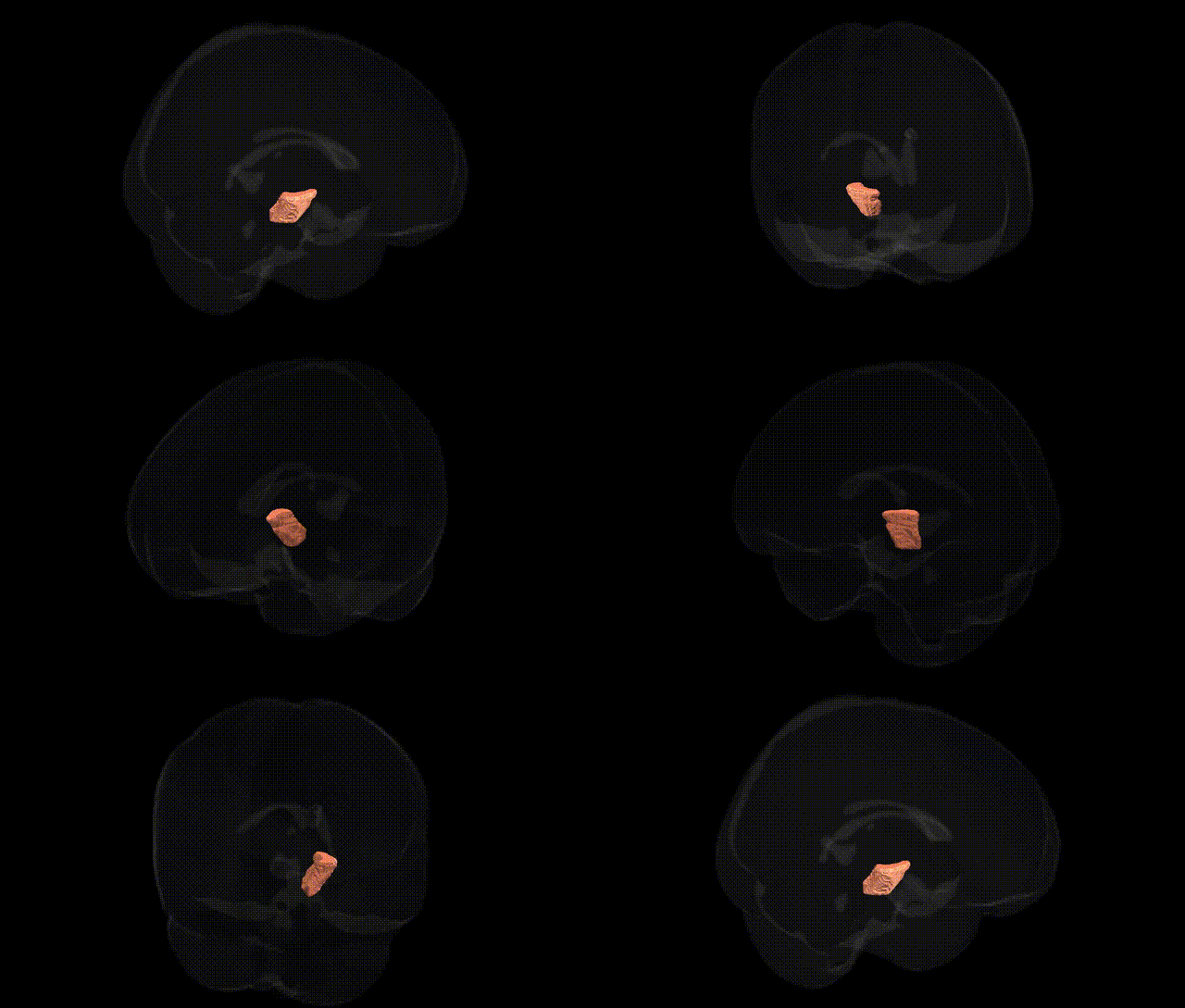
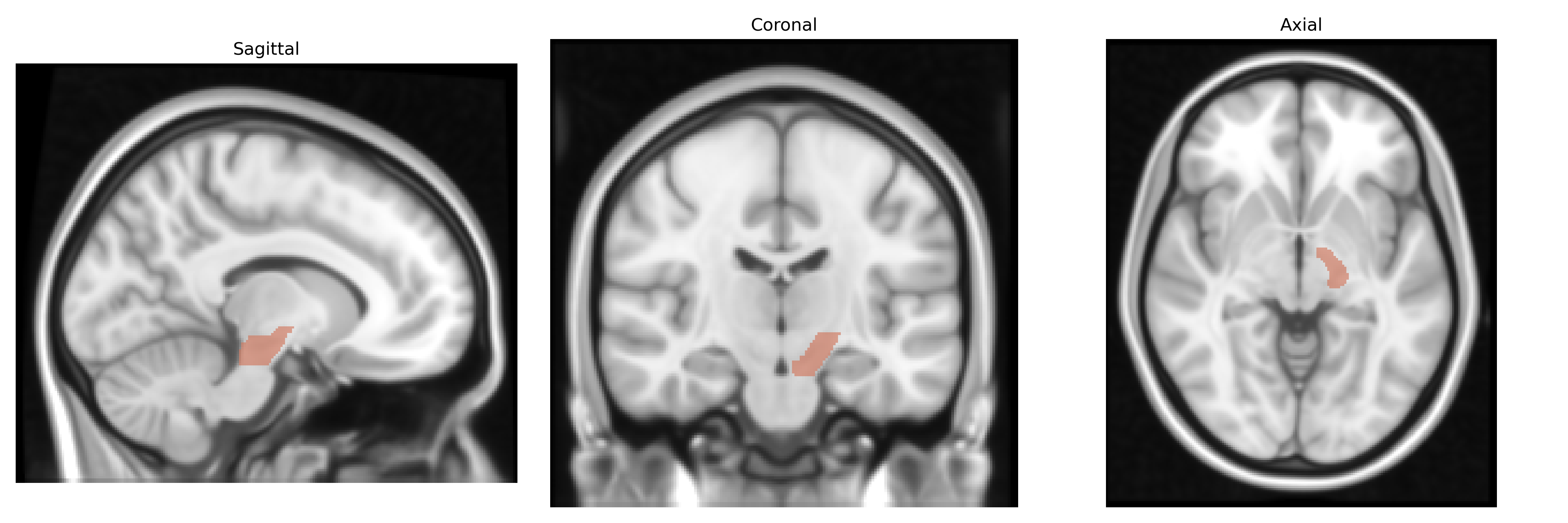
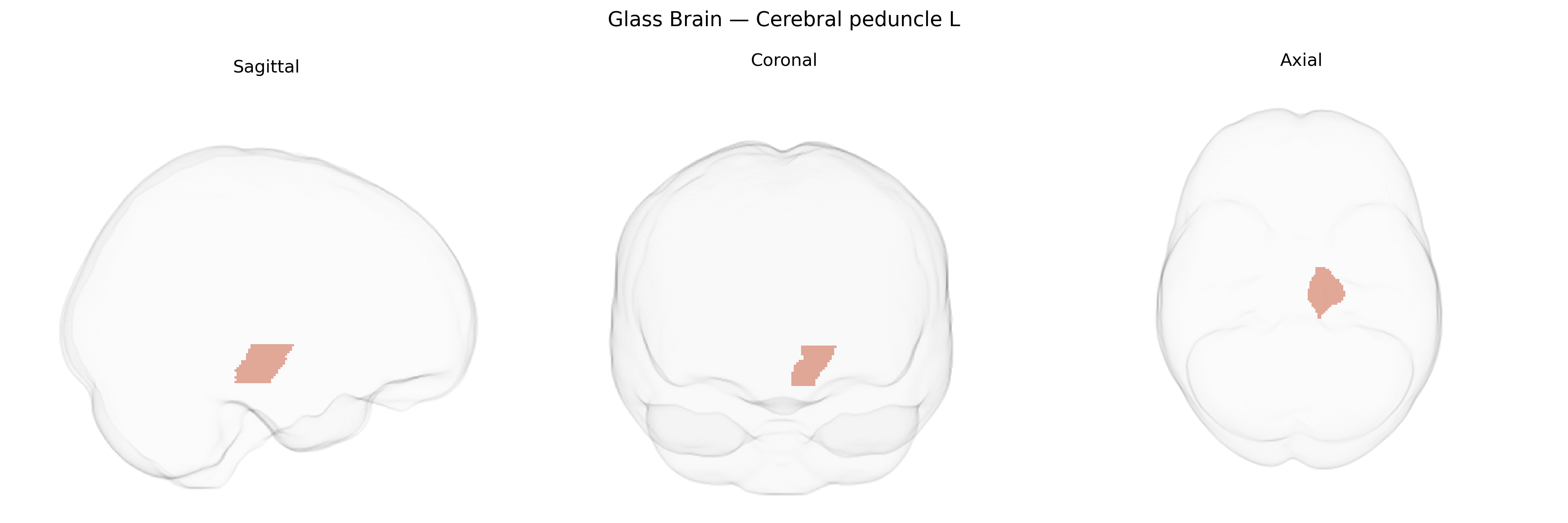
The bilateral cerebral peduncle (left) in the JHU ICBM 1 mm atlas corresponds to a major white matter structure in the midbrain that contains descending fiber tracts connecting the cerebral cortex to the brainstem and spinal cord. Composed predominantly of corticospinal, corticobulbar, and corticopontine fibers, the cerebral peduncle plays a critical role in voluntary motor control, modulation of reflexes, and integration of motor commands with subcortical and cerebellar circuits. Damage to this region can result in severe motor deficits such as contralateral hemiparesis, facial weakness, and disruption of motor planning and execution. There is no direct Wikipedia article for “Cerebral peduncle (left)”; a related structure encompassing this area is the Cerebral peduncle.
The bilateral cerebral peduncle (L) region defined in the JHU ICBM 1 mm atlas lies within the brainstem’s major descending motor pathways, but current GWAS and imaging-genetics literature rarely implicates this specific labeled region by name, and most genetic associations are reported at the level of white-matter tracts, global brainstem measures, or broader motor system structures. Large neuroimaging GWAS consortia such as ENIGMA and UK Biobank have identified variants in genes involved in myelination and axonal integrity (e.g., MAG, MBP, CNTN4, NRCAM, and loci near oligodendrocyte-related genes) that influence diffusion tensor imaging metrics across multiple white-matter regions, which plausibly include the cerebral peduncles, although tract-specific results are usually aggregated and not atlas-label specific. Genetic studies of disorders that prominently affect the cerebral peduncles, such as hereditary spastic paraplegias, leukodystrophies, and some spinocerebellar ataxias, have shown mutations in genes like SPAST, ATL1, PLP1, and others that cause degeneration or dysmyelination of corticospinal tracts traversing the peduncles, but these are typically described in clinical-neuroradiological terms rather than atlas-based regional GWAS hits. Imaging-genetic work in amyotrophic lateral sclerosis, multiple sclerosis, and cerebral small vessel disease has reported associations between polygenic risk and microstructural changes in brainstem motor pathways, yet again without singling out the JHU-defined cerebral peduncle L label. Overall, while numerous genetic variants are linked to white-matter microstructure and to diseases that affect the cerebral peduncles, no robust, widely replicated GWAS findings specifically target the bilateral cerebral peduncle (L) region as defined in the JHU ICBM labels 1 mm Atlas, and genetic associations remain largely inferred from broader tract or disease-based analyses rather than region-specific results.
Overview generated by GPT-4o (2026).
Region ID: 16
Hemisphere: bilateral
Atlas: JHU ICBM labels 1mm
Full Quality Version: Download MP4
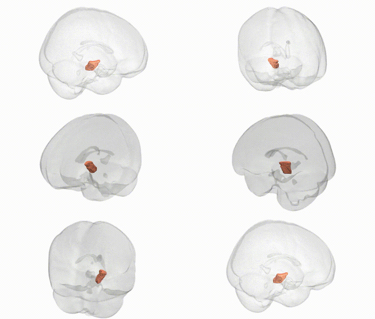
Full Quality Version: Download MP4
Wali Sidiqyar*, Gaurav Rudravaram*, Elyssa M. McMaster, Trent M. Schwartz, Adam M. Saunders, Kurt G. Schilling, Bennett A. Landman "Introducing SPINS: A Shared Public Visualization Library of Neuroanatomical Structures." Medical Imaging with Deep Learning- short paper
This resource is licensed under CC0 1.0 Universal (Public Domain).