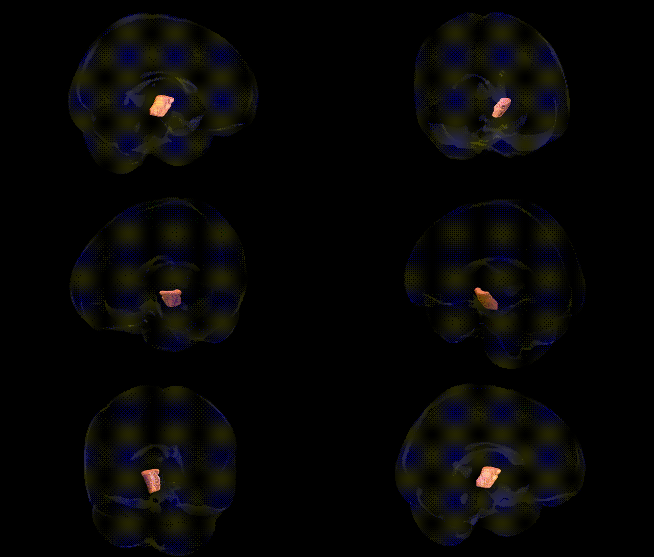
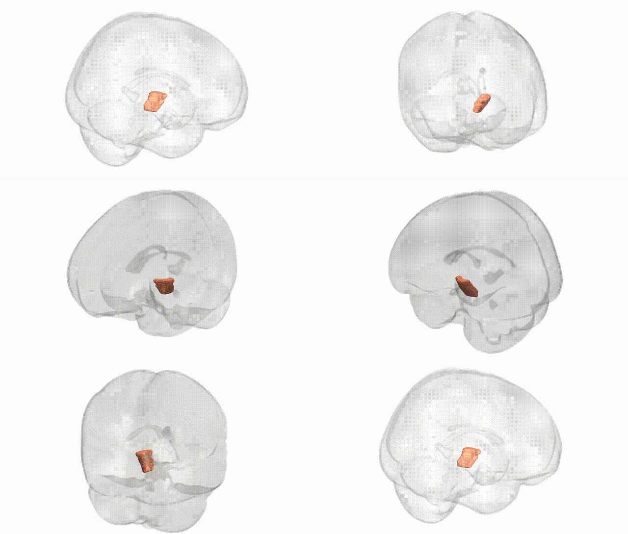
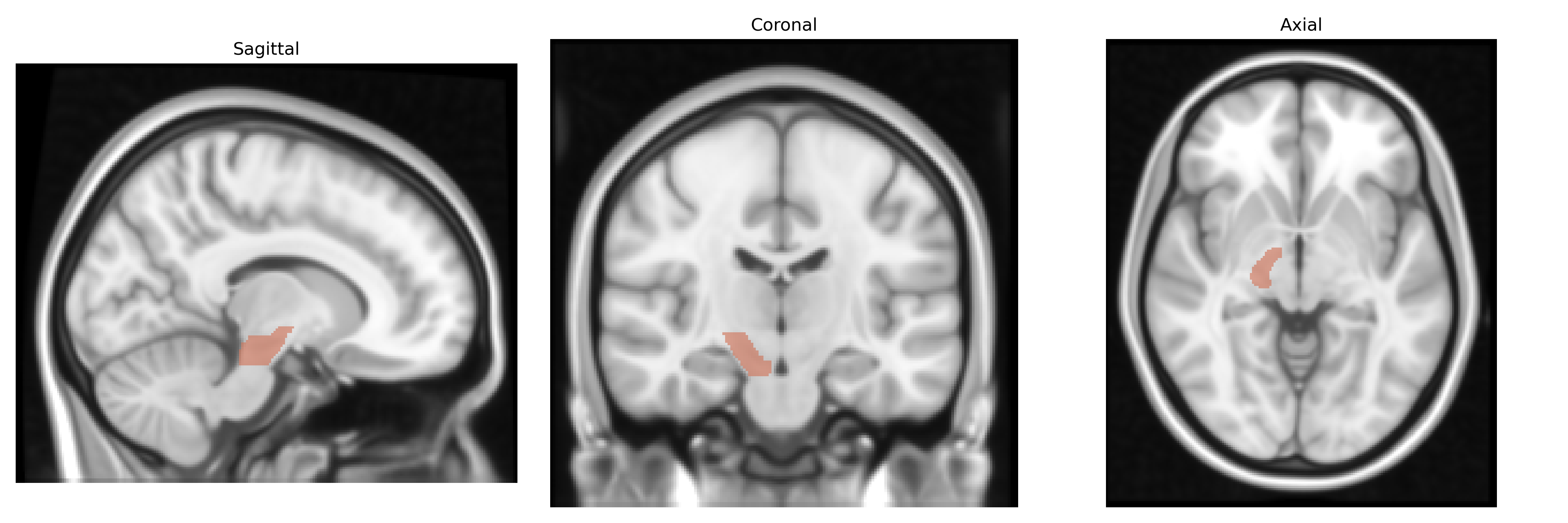
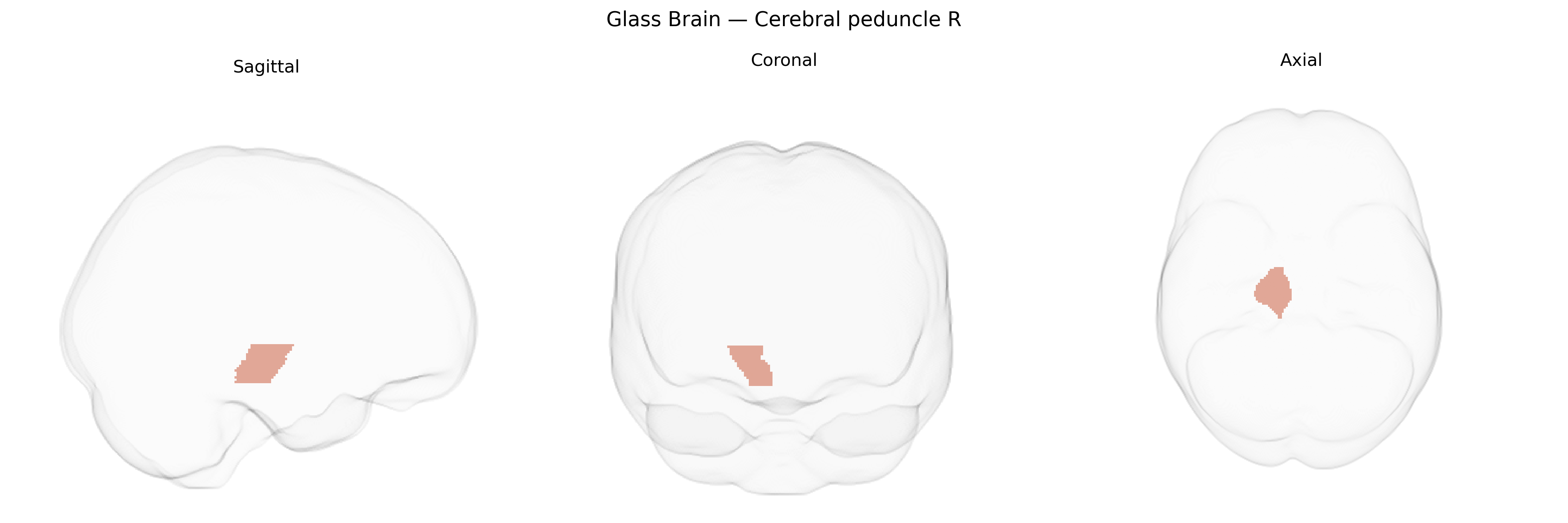
The bilateral cerebral peduncle (right) in the JHU ICBM 1 mm atlas corresponds to the right-sided portion of the cerebral peduncles, large white matter structures in the ventral midbrain that convey major descending motor pathways from the cerebral cortex to the brainstem and spinal cord. These peduncles contain the corticospinal, corticobulbar, and corticopontine tracts, organized somatotopically, and serve as a critical conduit linking frontal and other cortical regions with motor nuclei and spinal motor circuits. Damage to this region can produce contralateral motor deficits, including weakness, spasticity, and abnormalities of muscle tone, reflecting disruption of pyramidal and associated descending pathways. There is no direct Wikipedia article specifically for the “cerebral peduncle (right)”; a closely related structure is the midbrain: Midbrain.
Genetic associations specifically targeting the bilateral cerebral peduncle (right) as labeled in the JHU ICBM 1 mm atlas are limited, but several neuroimaging GWAS and diffusion tensor imaging (DTI) studies have implicated this white matter pathway and its microstructural properties (e.g., fractional anisotropy and mean diffusivity) in polygenic influences on motor, cognitive, and neuropsychiatric traits. Large-scale imaging genetics consortia such as ENIGMA and UK Biobank have identified heritable variation in cerebral peduncle integrity associated with common variants in genes involved in axon guidance, myelination, and neurodevelopment (e.g., loci near or within CNTN4, NRG1/NRG3, and oligodendrocyte-related genes), though often reported at the level of broader corticospinal or brainstem tracts rather than this specific label. GWAS of Parkinson’s disease, multiple sclerosis, and other movement or demyelinating disorders have linked risk variants to degeneration and microstructural alteration in corticospinal and brainstem pathways that include the cerebral peduncle, but these associations are typically reported for regional measures (brainstem or corticospinal tract integrity, motor network connectivity) rather than the JHU-defined “cerebral peduncle R” region alone. In psychiatric genetics, polygenic risk scores for schizophrenia, bipolar disorder, ADHD, and major depressive disorder have been correlated with altered white matter microstructure across motor and associative tracts, with some studies indicating subtle, genetically influenced differences in peduncular integrity, yet the findings are generally modest, nonspecific, and distributed across many regions. Overall, the cerebral peduncle is recognized as a heritable white matter structure whose microstructural measures are influenced by common genetic variation and by genetic risk for movement and psychiatric disorders, but current evidence treats it as part of broader corticospinal and brainstem networks, and no single locus or disorder is uniquely or definitively tied to the bilateral cerebral peduncle (right) region in existing GWAS.
Overview generated by GPT-4o (2026).
Region ID: 15
Hemisphere: bilateral
Atlas: JHU ICBM labels 1mm
Full Quality Version: Download MP4
Full Quality Version: Download MP4
Wali Sidiqyar*, Gaurav Rudravaram*, Elyssa M. McMaster, Trent M. Schwartz, Adam M. Saunders, Kurt G. Schilling, Bennett A. Landman "Introducing SPINS: A Shared Public Visualization Library of Neuroanatomical Structures." Medical Imaging with Deep Learning- short paper
This resource is licensed under CC0 1.0 Universal (Public Domain).