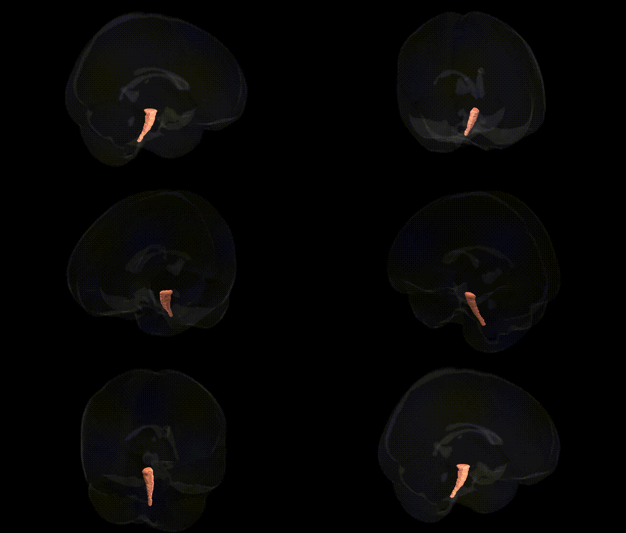
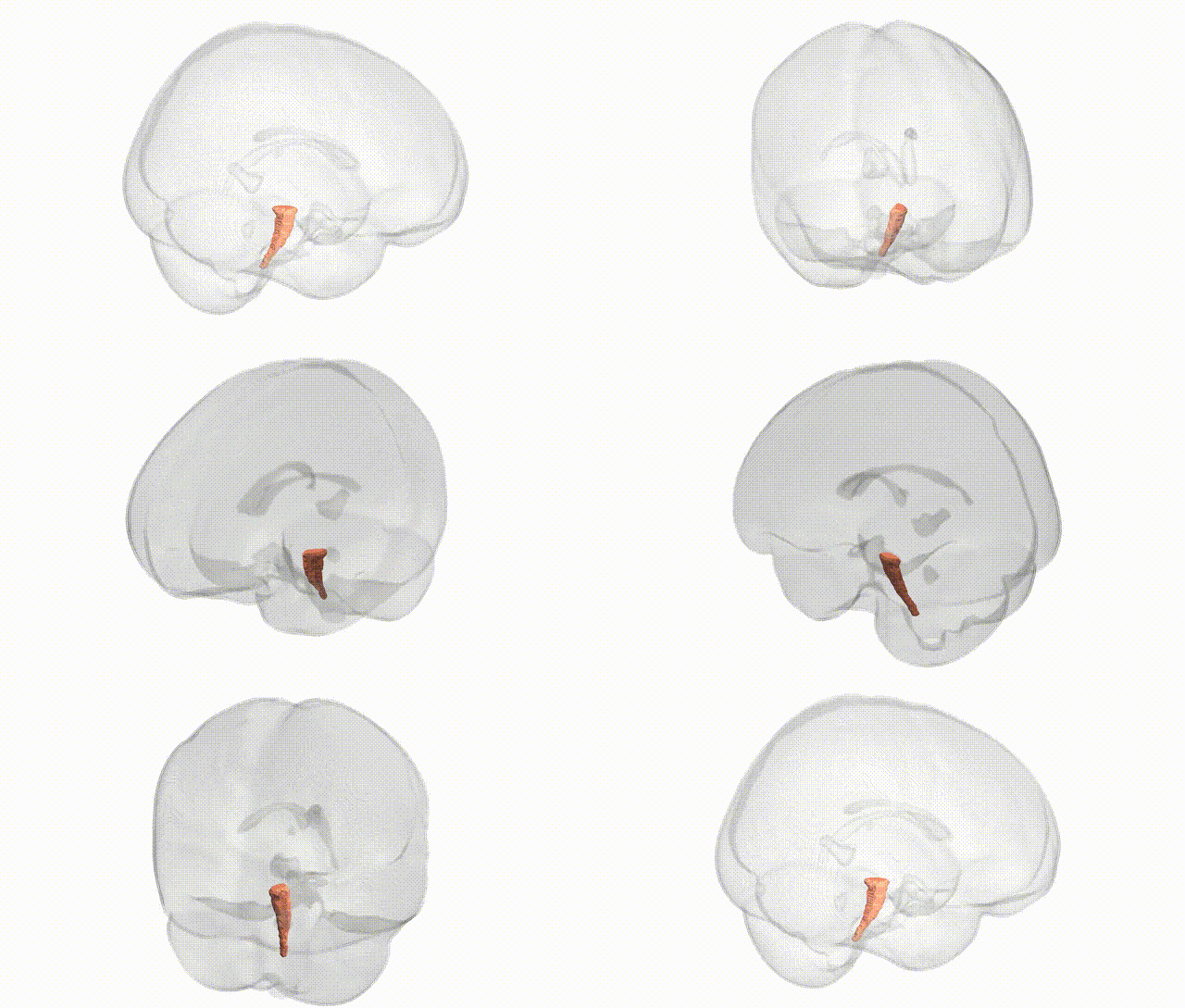
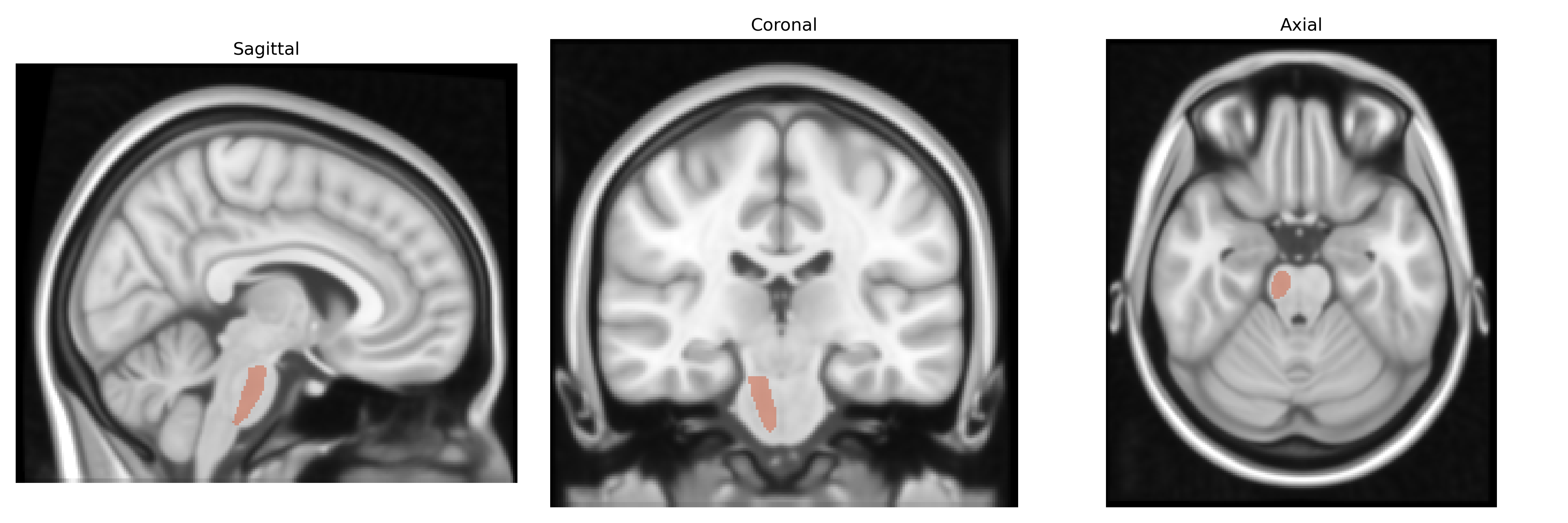
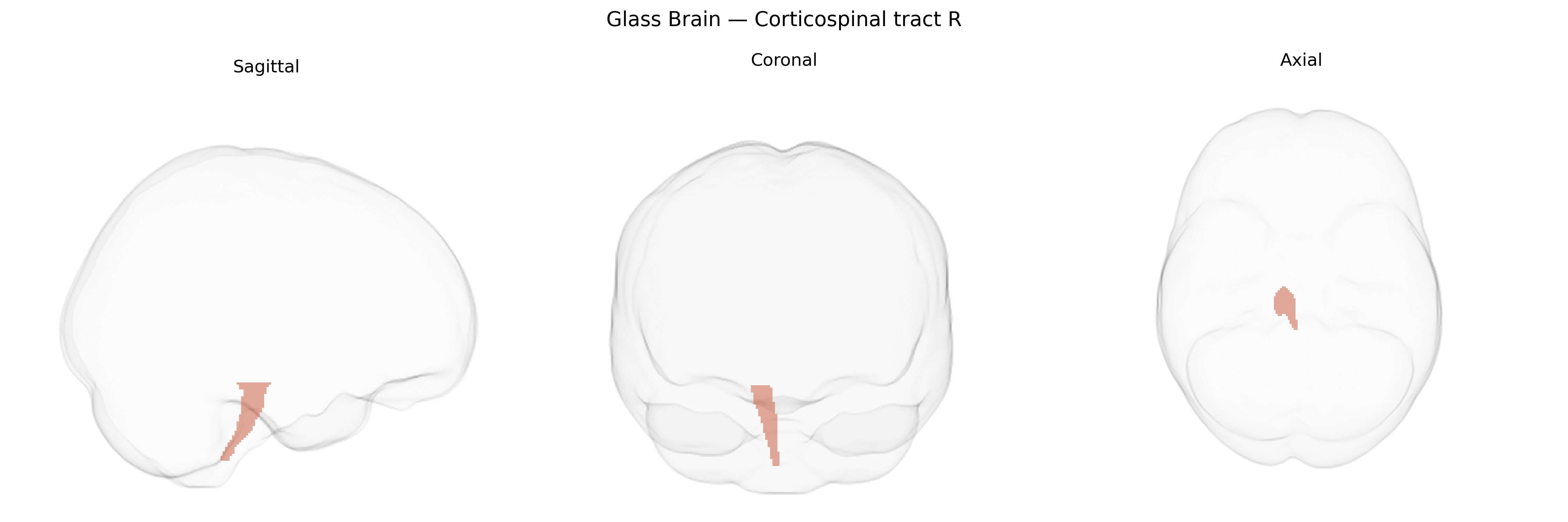
The bilateral corticospinal tract (right) in the JHU ICBM 1 mm atlas represents the major descending motor pathway conveying signals from the cerebral cortex to the spinal cord, primarily originating from pyramidal neurons in the primary motor cortex, premotor cortex, and supplementary motor areas. Fibers from both hemispheres descend through the corona radiata and internal capsule, traverse the cerebral peduncles, pass through the ventral pons, and form the medullary pyramids, where the majority decussate to influence contralateral spinal motor circuits, with a smaller proportion remaining ipsilateral. Functionally, this tract is crucial for the execution and fine control of voluntary movements, particularly of distal limb musculature, and damage to it can result in characteristic upper motor neuron signs such as weakness, spasticity, and hyperreflexia. There is no direct Wikipedia article specifically for the “bilateral corticospinal tract R” atlas label; a closely related structure is the corticospinal tract: Corticospinal tract.
The bilateral corticospinal tract, as defined in the JHU ICBM 1 mm atlas, has been implicated in genetic studies primarily through its role in motor function, neurodevelopment, and neurodegeneration, rather than through region-specific GWAS signals unique to this tract. Polygenic influences on corticospinal tract microstructure and integrity are reflected in diffusion MRI GWAS that identify associations between white matter metrics in motor pathways and common variants in genes involved in myelination, axon guidance, and neurodevelopment (for example, genes affecting oligodendrocyte function and neuronal growth cones), though these signals are typically reported for broader internal capsule or motor-related white matter regions rather than the corticospinal tract label alone. In disease-focused genetics, rare and common variants in ALS/primary lateral sclerosis–related genes (such as SOD1, C9orf72, FUS, TARDBP, and others) and hereditary spastic paraplegia genes (for example SPAST, ATL1, KIF5A, PLP1) are consistently associated with degeneration or thinning of the corticospinal tract on MRI or neuropathology, linking these genetic loci to structural changes in this pathway. Multiple sclerosis risk loci (including HLA region variants and non-HLA immune-related genes) have been associated with lesion burden and atrophy in motor tracts, including the corticospinal tract, in imaging–genetics analyses, while stroke and leukoaraiosis GWAS implicate vascular and blood–brain barrier genes that, when damaged, frequently affect this tract and thereby motor outcomes. Overall, existing genetic findings point to the corticospinal tract as a convergent target of genes regulating motor neuron integrity, white matter development, and immune-mediated demyelination, but there is limited evidence for genetic variants discovered explicitly and uniquely by GWAS of the JHU-defined bilateral corticospinal tract region itself.
Overview generated by GPT-4o (2026).
Region ID: 7
Hemisphere: bilateral
Atlas: JHU ICBM labels 1mm
Full Quality Version: Download MP4
Full Quality Version: Download MP4
Wali Sidiqyar*, Gaurav Rudravaram*, Elyssa M. McMaster, Trent M. Schwartz, Adam M. Saunders, Kurt G. Schilling, Bennett A. Landman "Introducing SPINS: A Shared Public Visualization Library of Neuroanatomical Structures." Medical Imaging with Deep Learning- short paper
This resource is licensed under CC0 1.0 Universal (Public Domain).