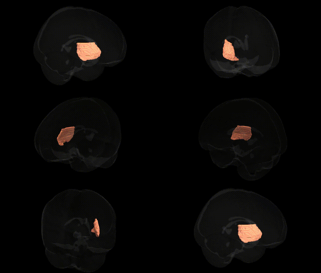
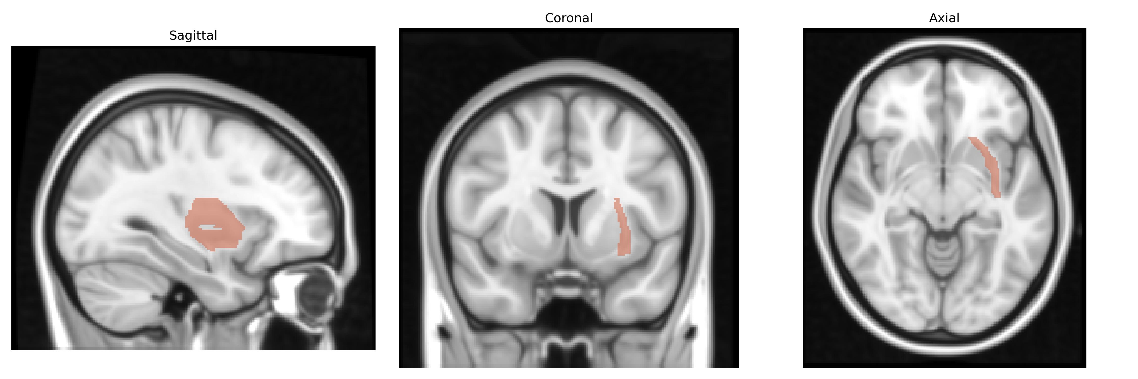
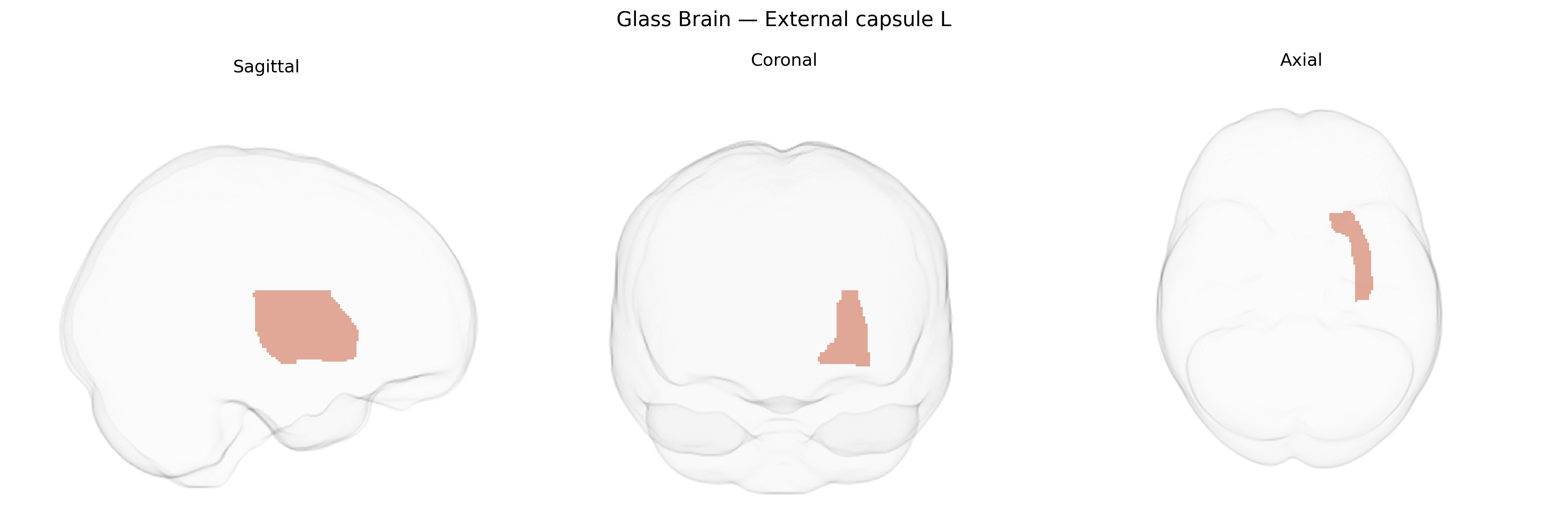
The bilateral external capsule is a sheet of white matter fibers located lateral to the putamen and medial to the claustrum, forming part of the subcortical connectivity system of the basal telencephalon. It contains association fibers such as components of the inferior fronto-occipital fasciculus and other cortico-cortical pathways that interconnect frontal, temporal, and occipital regions, and contributes to the integration of sensory, motor, and cognitive information. The external capsule is closely related to the internal capsule and extreme capsule, and lies within the broader basal ganglia–claustrum complex; its integrity is often assessed in diffusion MRI studies, including those based on the JHU ICBM white-matter atlas, to examine microstructural properties and network connectivity in health and disease. There is no direct Wikipedia article for the external capsule as an isolated entry; a closely related structure and context is the Internal capsule.
Genetic associations specific to the bilateral External capsule L region, as defined in the JHU ICBM labels 1 mm Atlas, are not well characterized, as most neuroimaging genetics and GWAS studies focus on larger white-matter tracts or global measures rather than this discrete structure. The external capsule contains association fibers connecting frontal, temporal, and limbic regions and is often subsumed within broader measures of fronto-temporal or limbic white matter in imaging genetics. Variants in genes involved in axon guidance, myelination, and synaptic plasticity (such as CNTNAP2, NRG1, and genes in the neuregulin–ErbB and glutamatergic pathways) have been repeatedly implicated in disorders that show microstructural changes in adjacent or overlapping tracts (e.g., schizophrenia, bipolar disorder, major depression, autism spectrum disorder), and some diffusion tensor imaging GWAS have linked common variants near genes involved in oligodendrocyte function (for example, regions near MAG, MBP, and other myelin-related loci) to white-matter integrity in fronto-temporal pathways that likely include fibers coursing through or near the external capsule. However, there are no widely replicated, region-specific GWAS findings tying particular SNPs or loci uniquely to external capsule L volume or diffusion metrics; existing evidence is largely indirect, arising from studies of broader white-matter networks and psychiatric or cognitive traits rather than targeted analyses of this atlas-defined region.
Overview generated by GPT-4o (2026).
Region ID: 34
Hemisphere: bilateral
Atlas: JHU ICBM labels 1mm
Full Quality Version: Download MP4
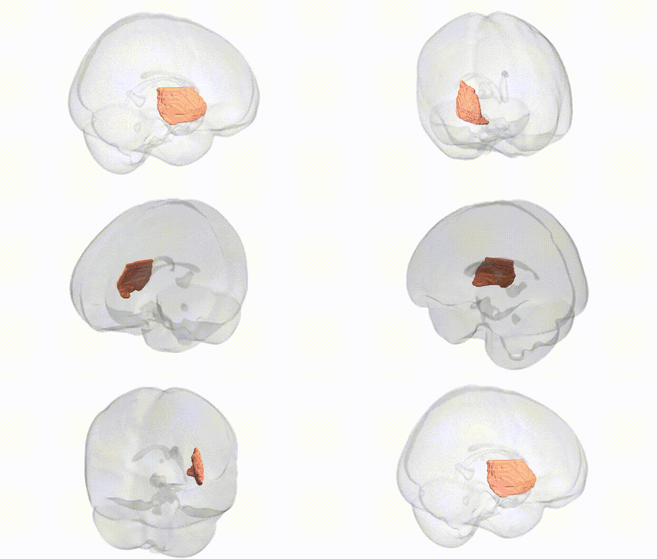
Full Quality Version: Download MP4
Wali Sidiqyar*, Gaurav Rudravaram*, Elyssa M. McMaster, Trent M. Schwartz, Adam M. Saunders, Kurt G. Schilling, Bennett A. Landman "Introducing SPINS: A Shared Public Visualization Library of Neuroanatomical Structures." Medical Imaging with Deep Learning- short paper
This resource is licensed under CC0 1.0 Universal (Public Domain).