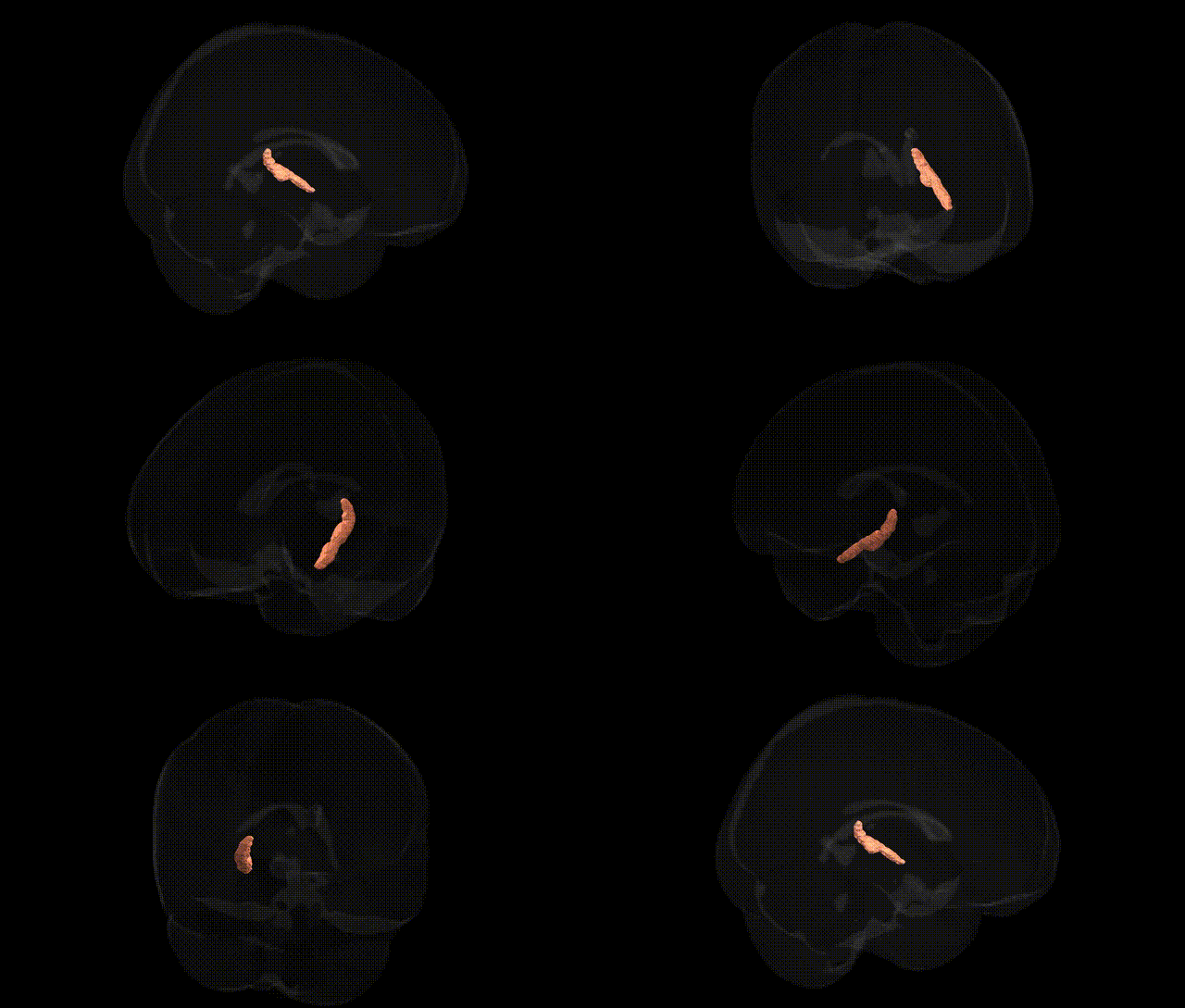
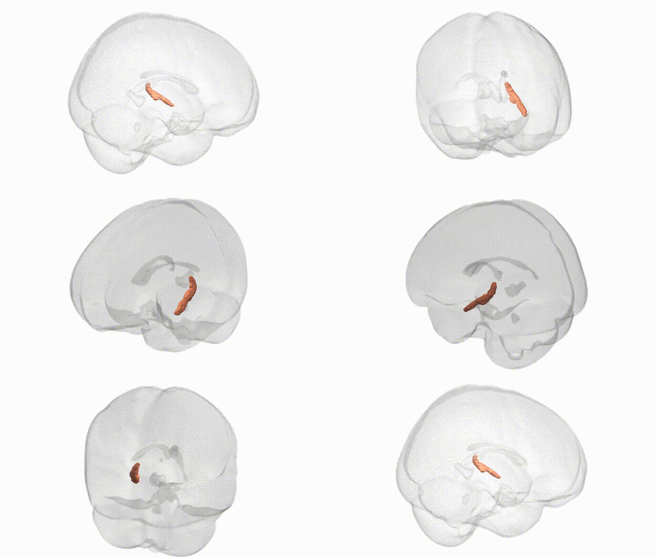
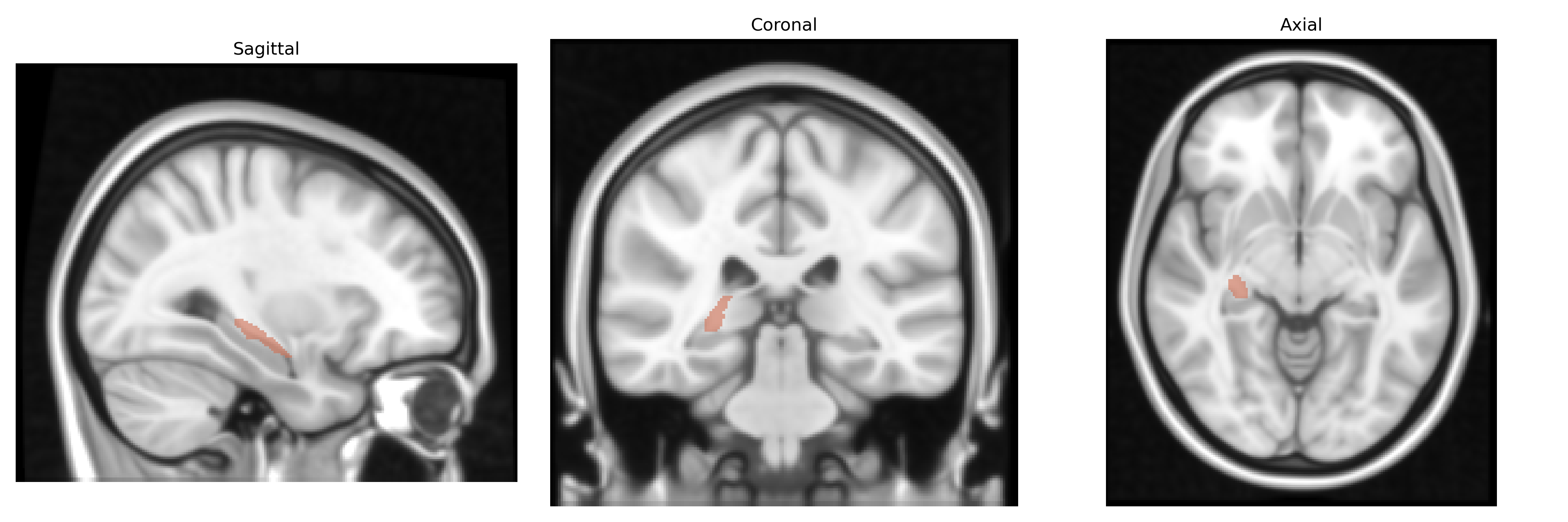
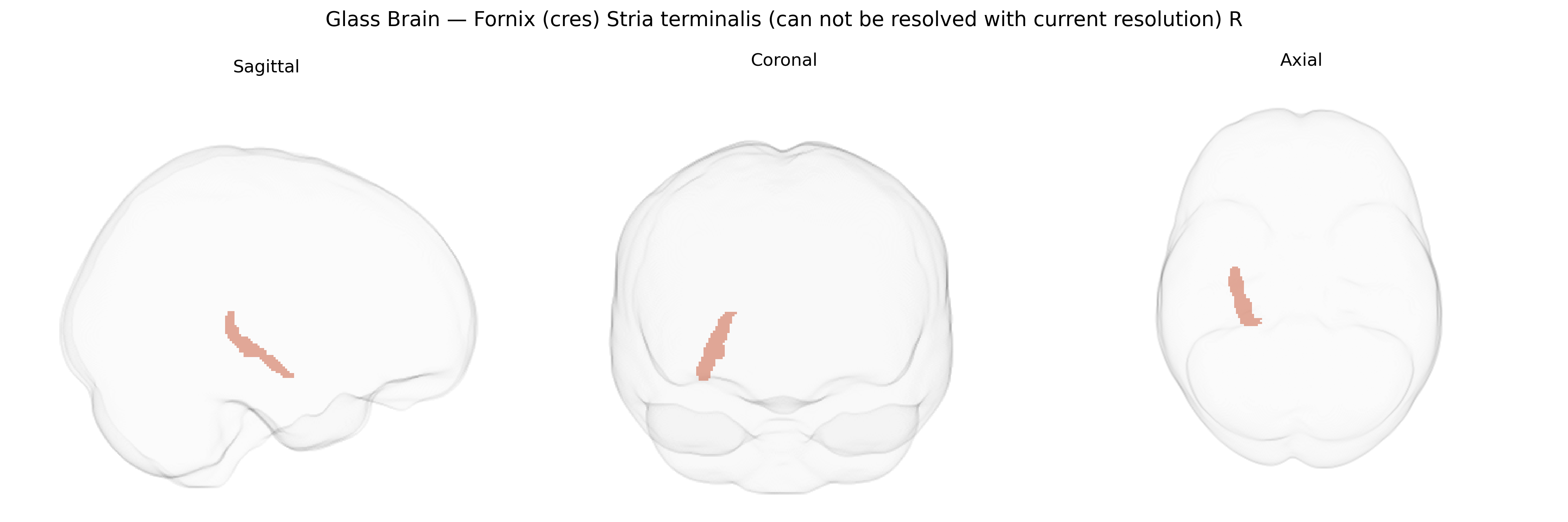
The bilateral fornix (cres) and stria terminalis are major white matter fiber bundles located deep within the medial temporal and diencephalic structures, forming key components of limbic circuitry. The fornix primarily carries efferent fibers from the hippocampus to the mammillary bodies and septal nuclei, supporting memory consolidation and aspects of spatial navigation. The stria terminalis conveys projections mainly from the amygdala to the hypothalamus and other basal forebrain structures, mediating neuroendocrine and autonomic responses associated with emotional and stress-related processing. In the JHU ICBM 1mm Atlas, these tracts are labeled together, reflecting their close anatomical proximity and overlapping roles in integrating mnemonic, emotional, and homeostatic functions, although they cannot be fully resolved at the given imaging resolution. There is no direct Wikipedia article for this combined label; a related entry is the Fornix (neuroanatomy).
The bilateral fornix (cres) and stria terminalis, as defined in the JHU ICBM 1 mm atlas, are key components of limbic circuitry, and while few GWAS directly target these specific labels, multiple genetic studies implicate them via related structural and functional phenotypes. Variants in genes involved in axon guidance, myelination, and synaptic plasticity—such as NRG1, CNTNAP2, and BDNF—have been associated with white matter integrity and diffusion tensor imaging (DTI) metrics in limbic tracts that include the fornix and stria terminalis. Large neuroimaging GWAS consortia (e.g., ENIGMA) have reported heritable variation in limbic white matter microstructure, with loci near genes like DAAM1, MAL, and others influencing fractional anisotropy and mean diffusivity in tracts connecting hippocampus, hypothalamus, and amygdala. Psychiatric and neurodevelopmental disorders characterized by limbic dysfunction, including schizophrenia, major depressive disorder, bipolar disorder, and post-traumatic stress disorder, show converging evidence of genetic risk (e.g., in CACNA1C, GRM3, SLC6A4) that is accompanied by alterations in fornix and stria terminalis connectivity or microstructure, although attribution to these specific atlas regions is indirect. Alzheimer’s disease and other dementias, strongly influenced by APOE and multiple GWAS loci, frequently demonstrate fornix degeneration, suggesting that genetic risk for neurodegeneration acts partly through vulnerability of these memory-related limbic pathways. Overall, genetic associations for this atlas-defined region emerge primarily from broader limbic tract and white matter imaging genetics rather than from studies focused exclusively on the “fornix (cres) stria terminalis (cannot be resolved with current resolution)” label.
Overview generated by GPT-4o (2026).
Region ID: 39
Hemisphere: bilateral
Atlas: JHU ICBM labels 1mm
Full Quality Version: Download MP4
Full Quality Version: Download MP4
Wali Sidiqyar*, Gaurav Rudravaram*, Elyssa M. McMaster, Trent M. Schwartz, Adam M. Saunders, Kurt G. Schilling, Bennett A. Landman "Introducing SPINS: A Shared Public Visualization Library of Neuroanatomical Structures." Medical Imaging with Deep Learning- short paper
This resource is licensed under CC0 1.0 Universal (Public Domain).