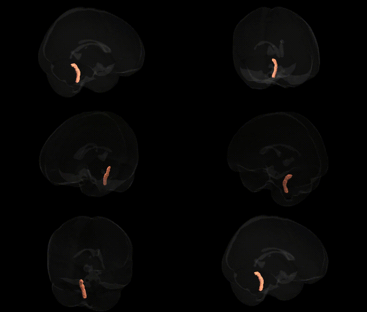
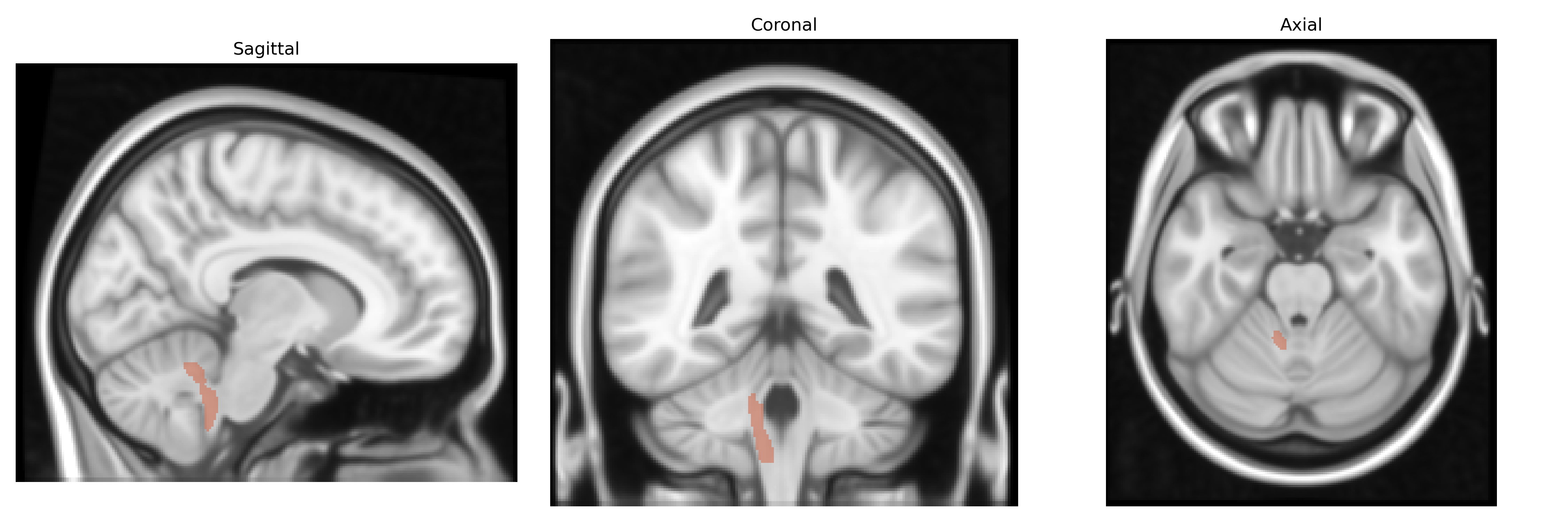
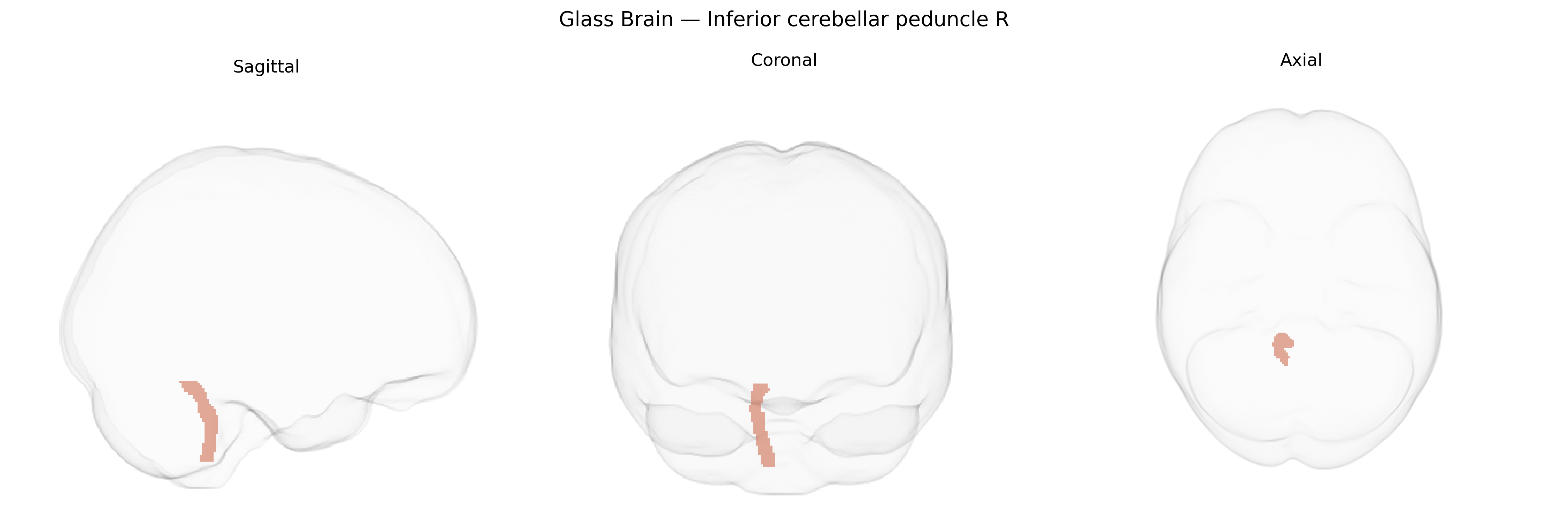
The bilateral Inferior cerebellar peduncle (right) is a major white matter tract that connects the medulla oblongata and spinal cord with the cerebellum, carrying predominantly afferent fibers that convey somatosensory, proprioceptive, and vestibular information essential for coordination, balance, and posture. It includes pathways such as the dorsal spinocerebellar tract, cuneocerebellar fibers, olivocerebellar fibers from the inferior olivary nucleus, and vestibulocerebellar inputs, which project mainly to the vermis and intermediate zones of the cerebellar cortex and deep nuclei. Functionally, this pathway supports fine-tuning of motor activity and adaptive motor learning by continuously updating cerebellar circuits with peripheral and brainstem sensory signals. Lesions involving the inferior cerebellar peduncle can lead to ipsilateral limb ataxia, dysmetria, gait disturbances, and vertigo due to disruption of cerebellar integration of sensory input. There is no direct Wikipedia article for the Inferior cerebellar peduncle; a related structure is the Cerebellar peduncle.
Genetic association data specifically targeting the bilateral inferior cerebellar peduncle (right) as labeled in the JHU ICBM 1 mm atlas are limited, but several large-scale imaging genetics and GWAS studies of white matter microstructure and cerebellar pathways implicate this region indirectly via diffusion tensor imaging (DTI) metrics such as fractional anisotropy and mean diffusivity. Variants in genes involved in axonal guidance, myelination, and cerebellar development—such as those affecting oligodendrocyte function (e.g., loci near MAG, NRG1, and other myelin-related genes) and axon growth (e.g., ROBO/SLIT pathway genes)—have been associated with global or regional white matter integrity that includes the inferior cerebellar peduncle. Polygenic influences on cerebellar and brainstem connectivity identified in ENIGMA and UK Biobank–based GWAS of DTI tracts extend to this pathway, although most reports aggregate across multiple cerebellar peduncles or brainstem tracts rather than isolating the right inferior peduncle. Clinically, genetic variants linked to spinocerebellar ataxias (e.g., repeat expansions in ATXN genes), leukodystrophies, and congenital cerebellar malformations often manifest radiologically with involvement of the inferior cerebellar peduncle, but such findings typically reflect broader cerebellar or brainstem pathology rather than tract-specific GWAS hits. Overall, current evidence supports polygenic, nonspecific contributions to inferior cerebellar peduncle structure and function within the general framework of white matter and cerebellar connectivity genetics, with no robust, widely replicated SNP associations uniquely mapped to this tract in the JHU ICBM atlas.
Overview generated by GPT-4o (2026).
Region ID: 11
Hemisphere: bilateral
Atlas: JHU ICBM labels 1mm
Full Quality Version: Download MP4
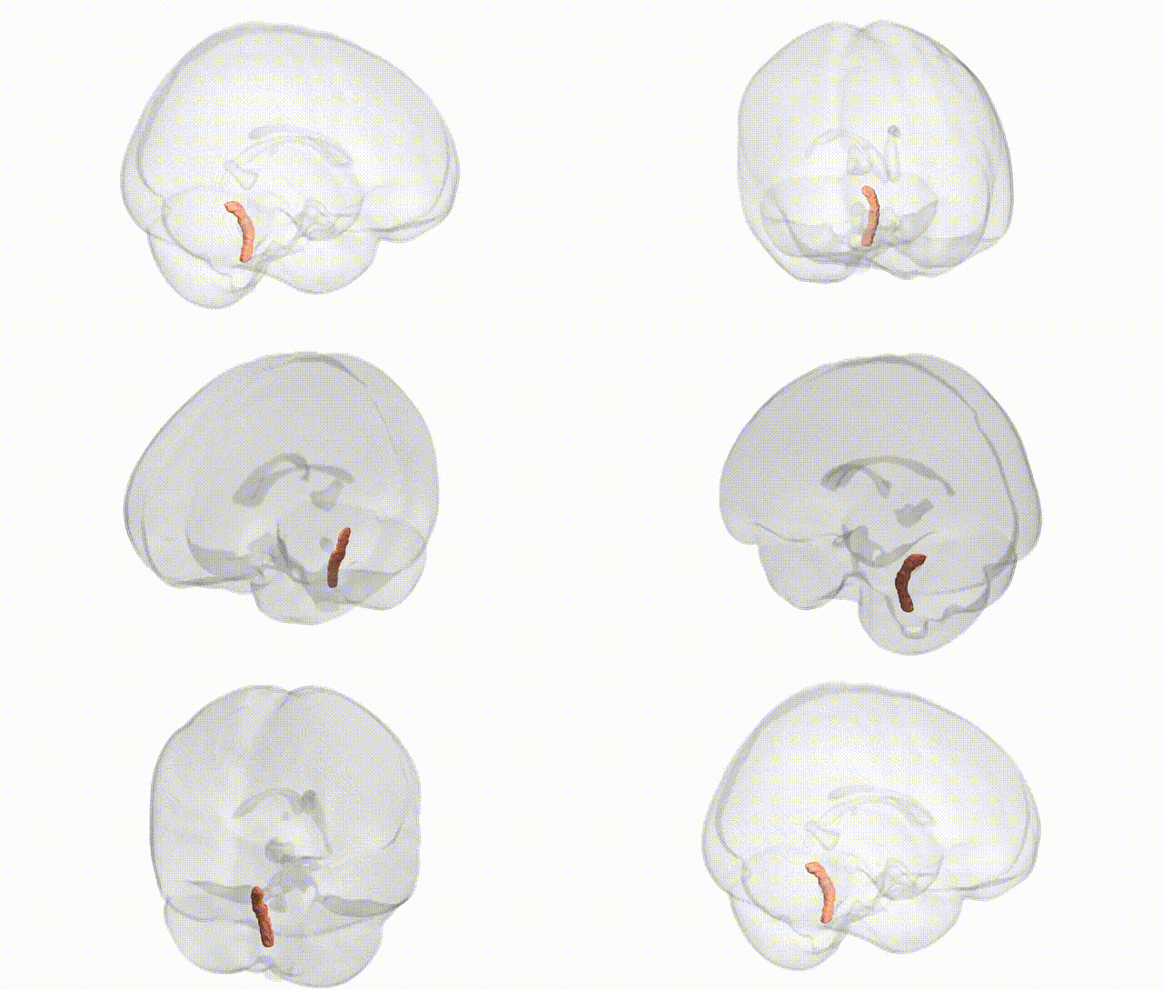
Full Quality Version: Download MP4
Wali Sidiqyar*, Gaurav Rudravaram*, Elyssa M. McMaster, Trent M. Schwartz, Adam M. Saunders, Kurt G. Schilling, Bennett A. Landman "Introducing SPINS: A Shared Public Visualization Library of Neuroanatomical Structures." Medical Imaging with Deep Learning- short paper
This resource is licensed under CC0 1.0 Universal (Public Domain).