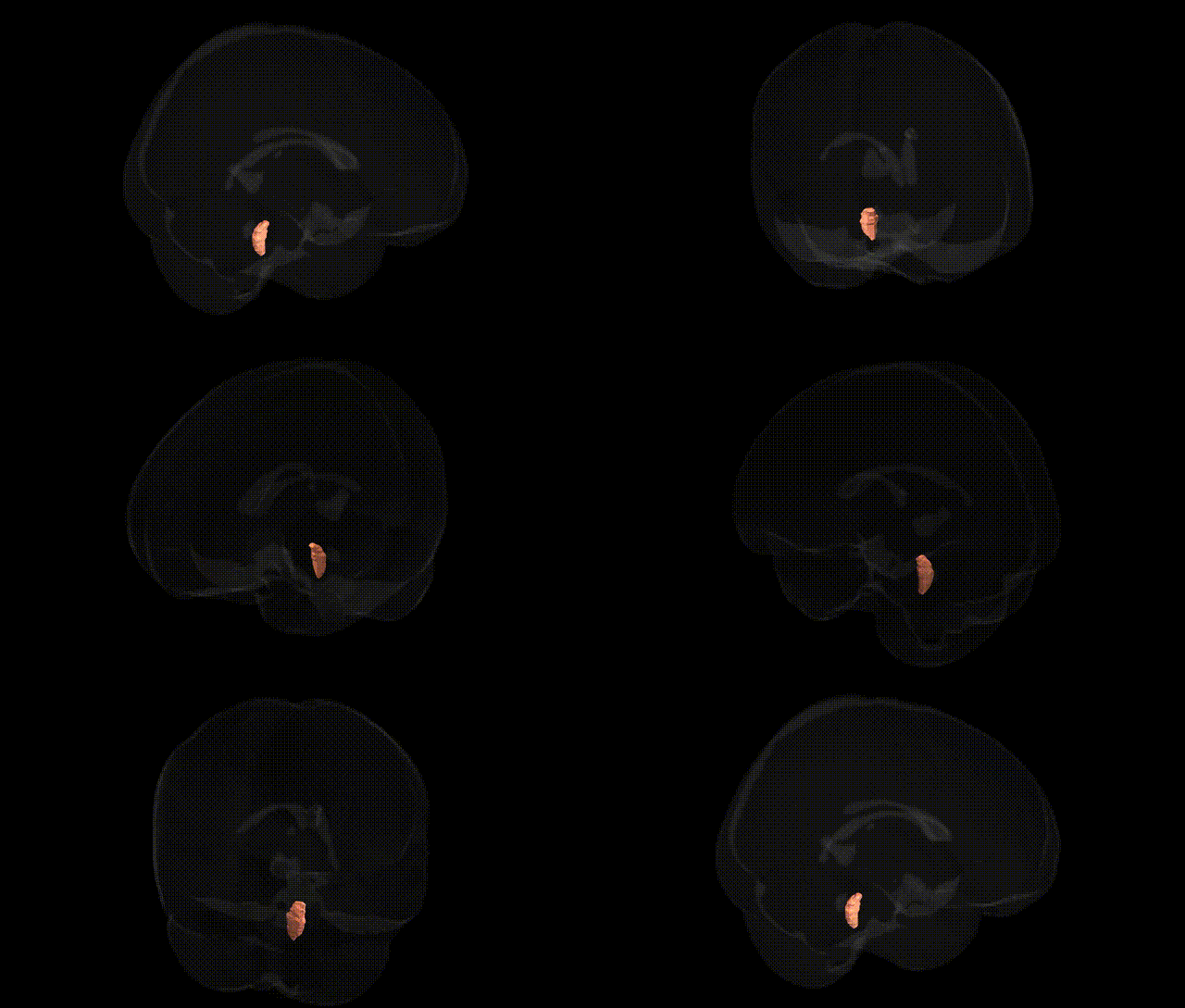
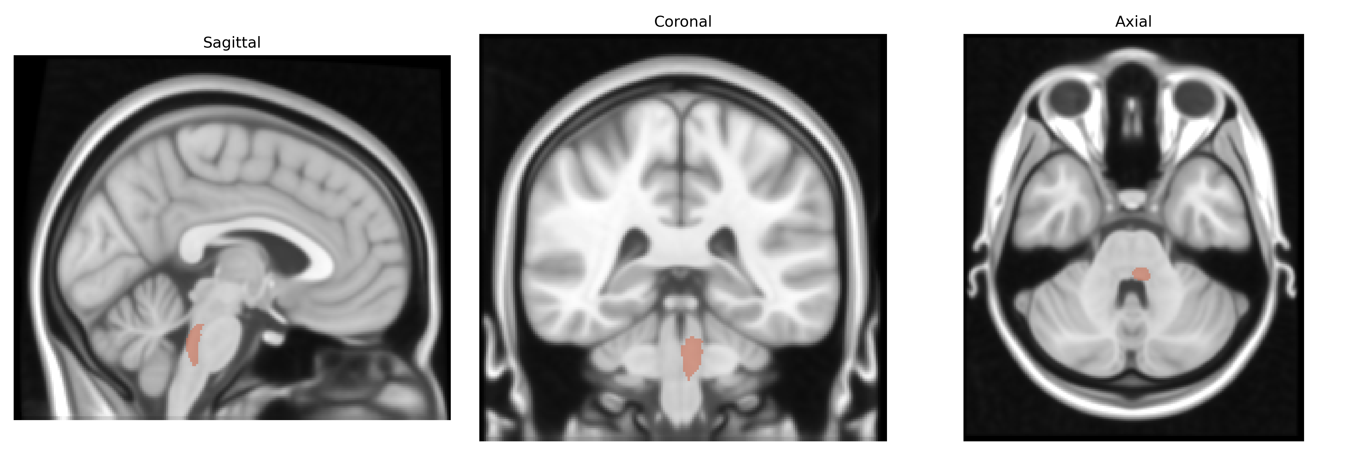
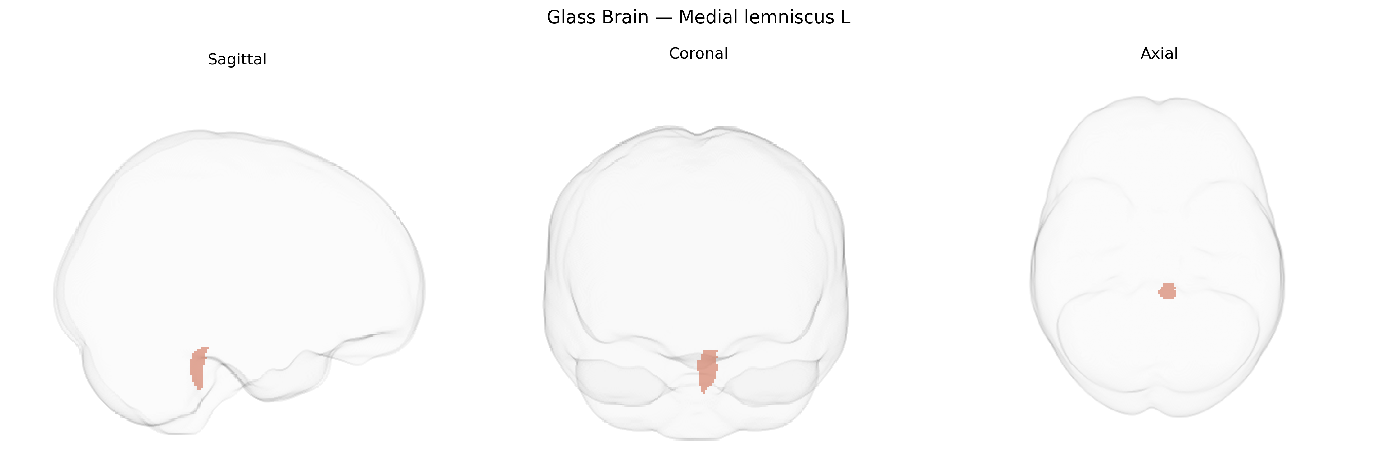
The bilateral medial lemniscus L, as defined in the JHU ICBM labels 1mm Atlas, corresponds to the left-sided portion of the medial lemniscus, a prominent ascending somatosensory pathway in the brainstem. It is formed by second-order neurons originating from the gracile and cuneate nuclei in the dorsal column nuclei of the medulla, which decussate as internal arcuate fibers and ascend through the brainstem to terminate in the ventral posterior nucleus of the thalamus. Functionally, this tract conveys fine touch, vibration, and proprioceptive information from the contralateral side of the body to higher cortical centers, contributing critically to discriminative somatosensory processing. No direct Wikipedia article exists for the atlas-defined “bilateral medial lemniscus L” label, but the structure is part of the medial lemniscus pathway: Medial lemniscus.
The bilateral medial lemniscus (L) region, as defined in the JHU ICBM labels 1 mm atlas, has relatively sparse direct genetic association data, as most GWAS and imaging genetics studies focus on cortical and large subcortical structures rather than specific brainstem tracts; however, several lines of evidence implicate genes influencing white matter microstructure and sensorimotor pathways in variation affecting this region. Diffusion MRI GWAS examining global and tract-specific white matter integrity have identified common variants in genes related to axon guidance, myelination, and neural development (such as CNTN4, NRG1, and MAG) that broadly impact brainstem and thalamocortical tracts, which likely include or intersect the medial lemniscus. Disorders with prominent involvement of dorsal column–medial lemniscal pathways—such as hereditary spastic paraplegias, certain spinocerebellar ataxias, and leukodystrophies—are linked to mutations in genes governing long-tract neuron maintenance and myelin (e.g., SPAST, ATL1, PLP1), and postmortem or advanced imaging studies in these conditions often demonstrate degeneration or altered integrity in medial lemniscus-related pathways. Large neuroimaging genetic consortia (e.g., ENIGMA) have reported polygenic influences on brainstem and global white matter diffusion metrics correlated with traits such as general cognitive ability, neuroticism, and risk for major depressive disorder and schizophrenia, suggesting that the medial lemniscus, as part of the broader sensory white matter scaffold, may be indirectly affected by polygenic architectures shaping white matter organization. Additionally, rare Mendelian sensory neuropathies and neurodevelopmental syndromes involving genes such as SLC12A5 and PMP22 can alter somatosensory transmission and brainstem sensory nuclei, implicating the lemniscal system functionally, although not typically isolating the medial lemniscus tract in genetic association maps. Overall, current genetic findings point to indirect, system-level influences on medial lemniscus structure and function via genes controlling axonal structure, myelination, and sensorimotor network development, while direct GWAS hits specifically annotated to the bilateral medial lemniscus (L) label in the JHU ICBM atlas remain limited or unreported in the literature to date.
Overview generated by GPT-4o (2026).
Region ID: 10
Hemisphere: bilateral
Atlas: JHU ICBM labels 1mm
Full Quality Version: Download MP4
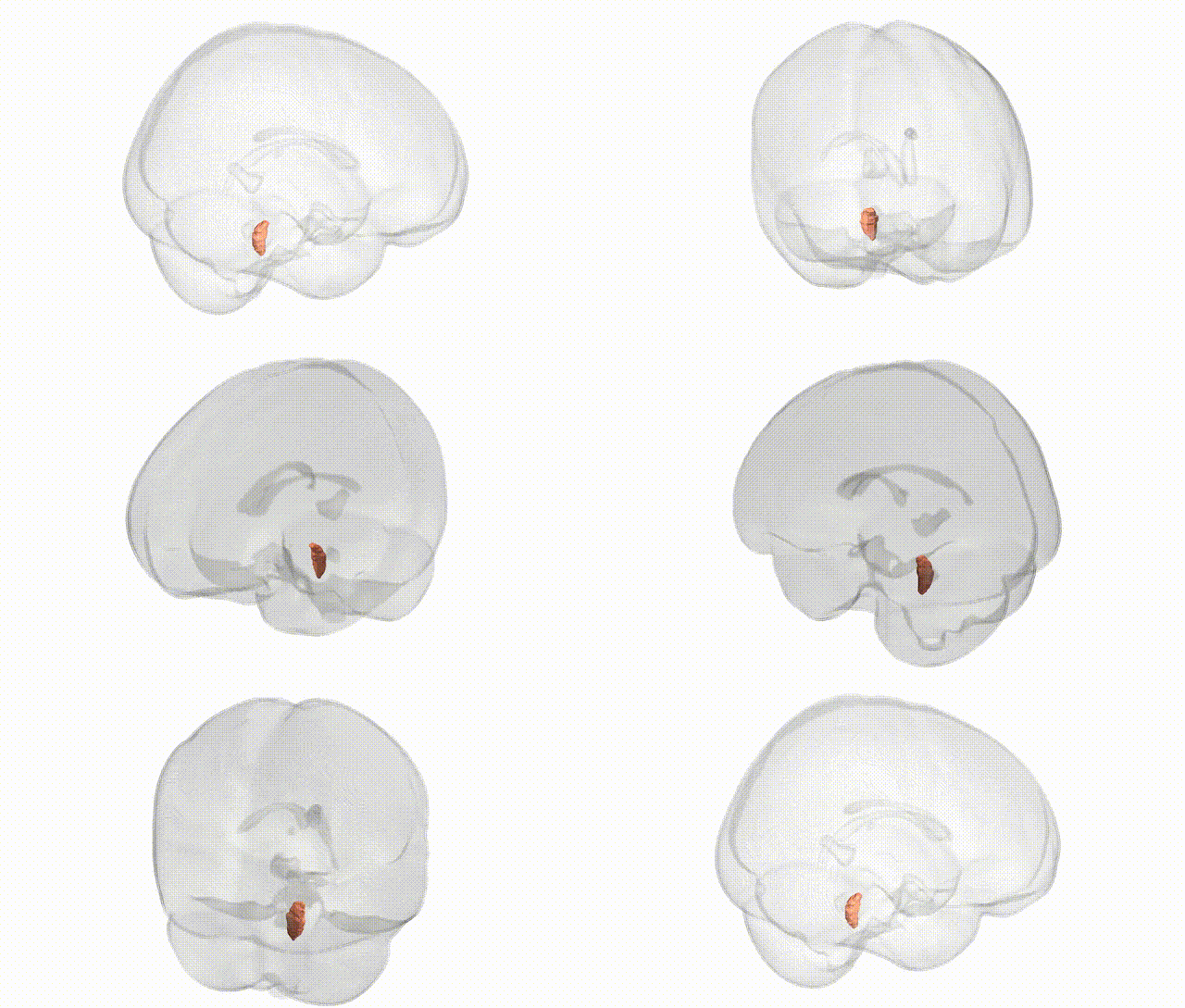
Full Quality Version: Download MP4
Wali Sidiqyar*, Gaurav Rudravaram*, Elyssa M. McMaster, Trent M. Schwartz, Adam M. Saunders, Kurt G. Schilling, Bennett A. Landman "Introducing SPINS: A Shared Public Visualization Library of Neuroanatomical Structures." Medical Imaging with Deep Learning- short paper
This resource is licensed under CC0 1.0 Universal (Public Domain).