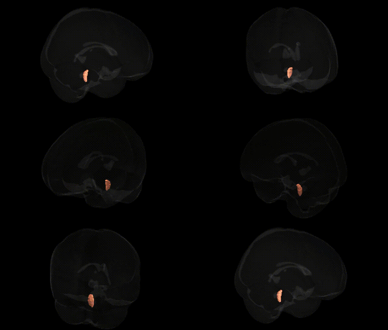
The bilateral medial lemniscus (right) is a major ascending white matter tract in the brainstem that conveys fine touch, vibration, and proprioceptive information from the contralateral side of the body to higher-order somatosensory structures. Arising from second-order neurons in the dorsal column nuclei (gracile and cuneate nuclei) after decussation in the medulla, its fibers ascend through the medulla, pons, and midbrain in a somatotopically organized manner, projecting primarily to the ventral posterior nucleus of the thalamus and subsequently to the primary somatosensory cortex. In diffusion MRI–based atlases such as the JHU ICBM 1 mm atlas, this tract is segmented bilaterally, with the “R” designation indicating the portion located in the right brainstem and associated with sensory input originating from the left side of the body. Medial lemniscus
The bilateral medial lemniscus, as defined in the JHU ICBM labels 1 mm atlas, has not been a primary focus of region-specific GWAS, but genetic associations implicating this brainstem sensory pathway arise indirectly through studies of white-matter microstructure, brainstem integrity, and disorders that affect dorsal column–medial lemniscus function. Large-scale imaging–genetics consortia (e.g., ENIGMA) and diffusion tensor imaging GWAS have linked common variants in genes related to myelination, axon guidance, and neurodevelopment—such as those in oligodendrocyte and cell-adhesion pathways—to interindividual differences in white-matter tracts that include or approximate brainstem sensory pathways, though results are typically reported at the tract or whole-brain level rather than specifically for the medial lemniscus. Clinically, rare mutations causing hereditary spastic paraplegias, leukodystrophies, and other neurodegenerative or demyelinating disorders (e.g., variants in PLP1, SPAST, GJC2, and related genes) can lead to degeneration or dysfunction of dorsal column–medial lemniscus structures, manifesting as impaired vibration and proprioceptive sensation; however, these findings reflect broader pathway involvement rather than a unique genetic signature for the medial lemniscus itself. Overall, genetic influences on this region are inferred from broader white-matter and neurodevelopmental genetic architectures rather than from medial-lemniscus–specific GWAS signals.
Overview generated by GPT-4o (2026).
Region ID: 9
Hemisphere: bilateral
Atlas: JHU ICBM labels 1mm
Full Quality Version: Download MP4
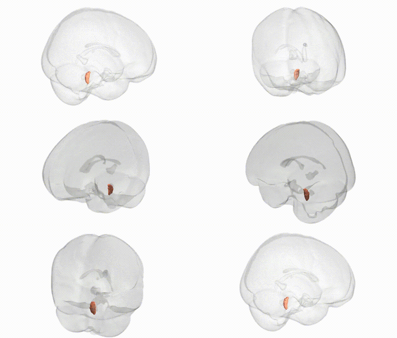
Full Quality Version: Download MP4
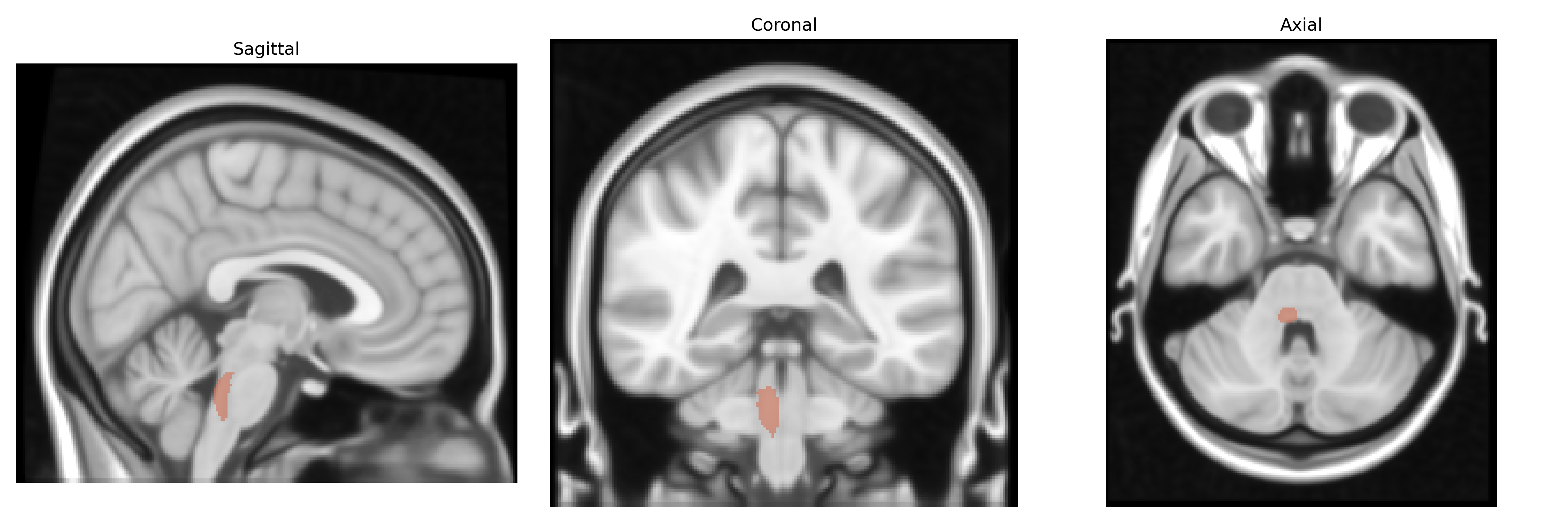
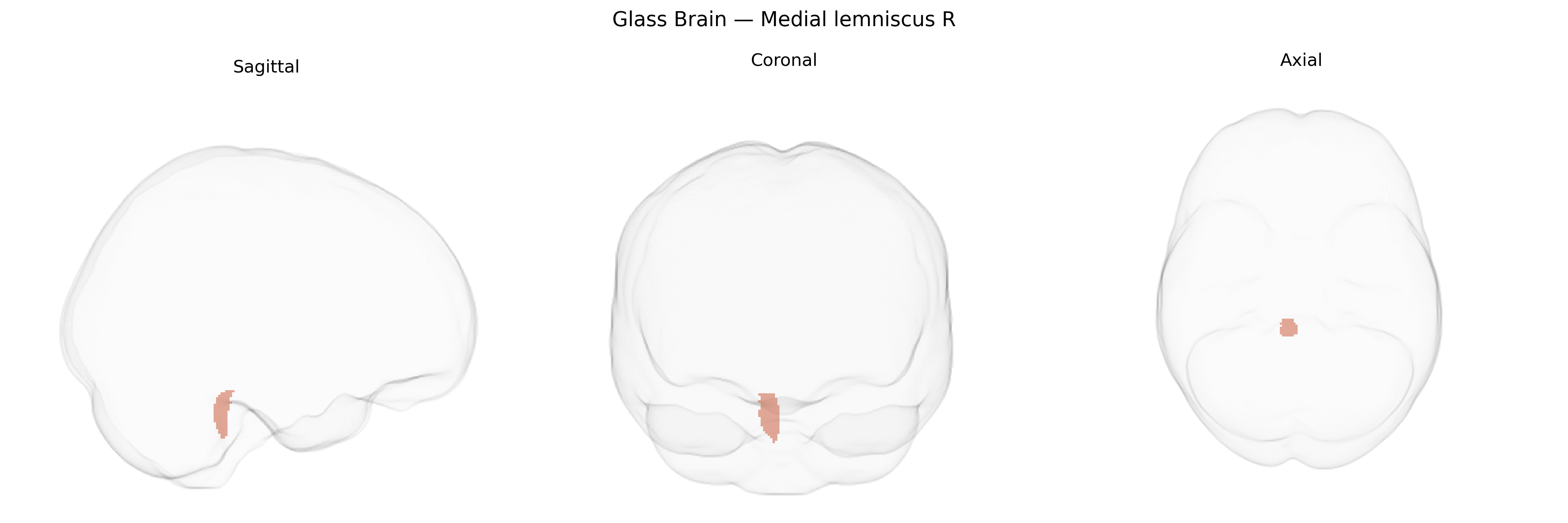
Wali Sidiqyar*, Gaurav Rudravaram*, Elyssa M. McMaster, Trent M. Schwartz, Adam M. Saunders, Kurt G. Schilling, Bennett A. Landman "Introducing SPINS: A Shared Public Visualization Library of Neuroanatomical Structures." Medical Imaging with Deep Learning- short paper
This resource is licensed under CC0 1.0 Universal (Public Domain).