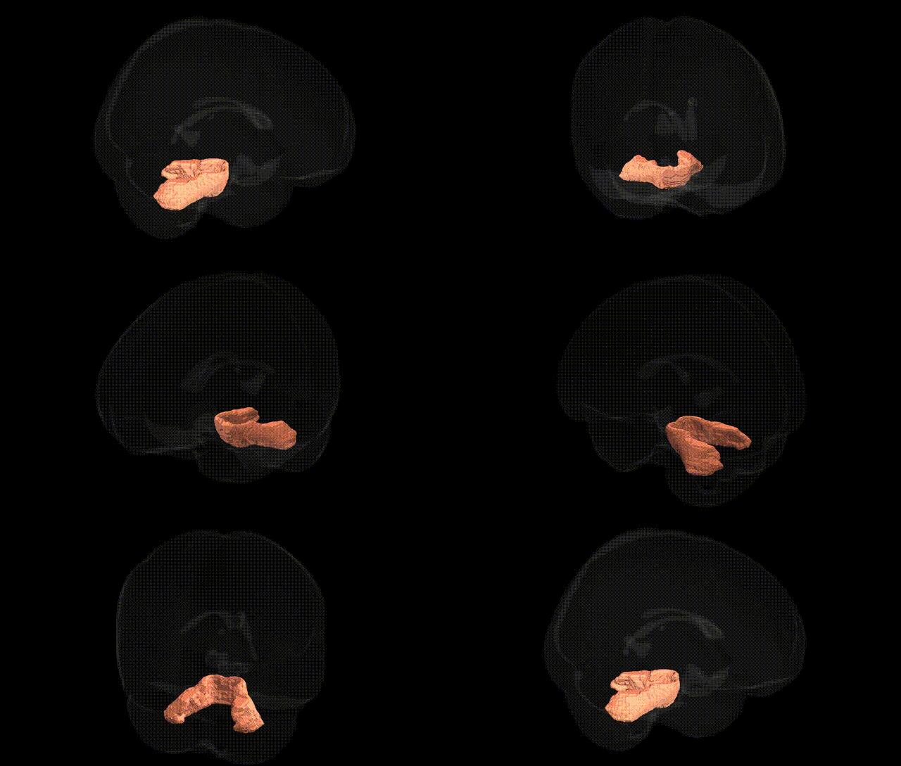
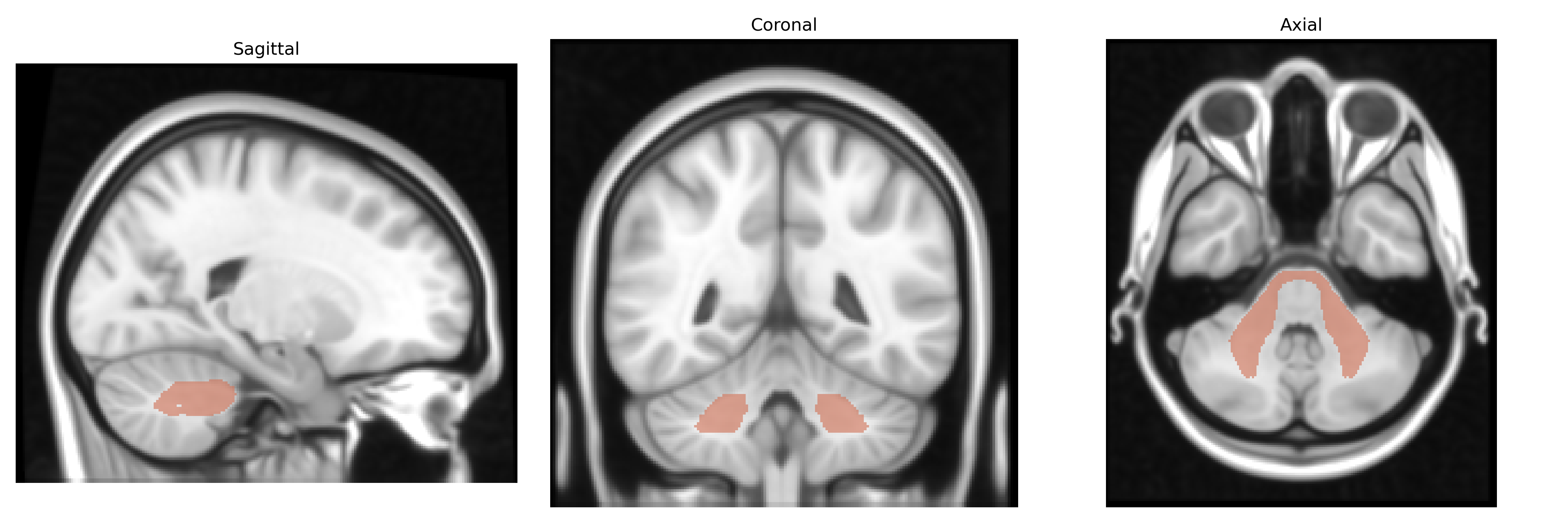
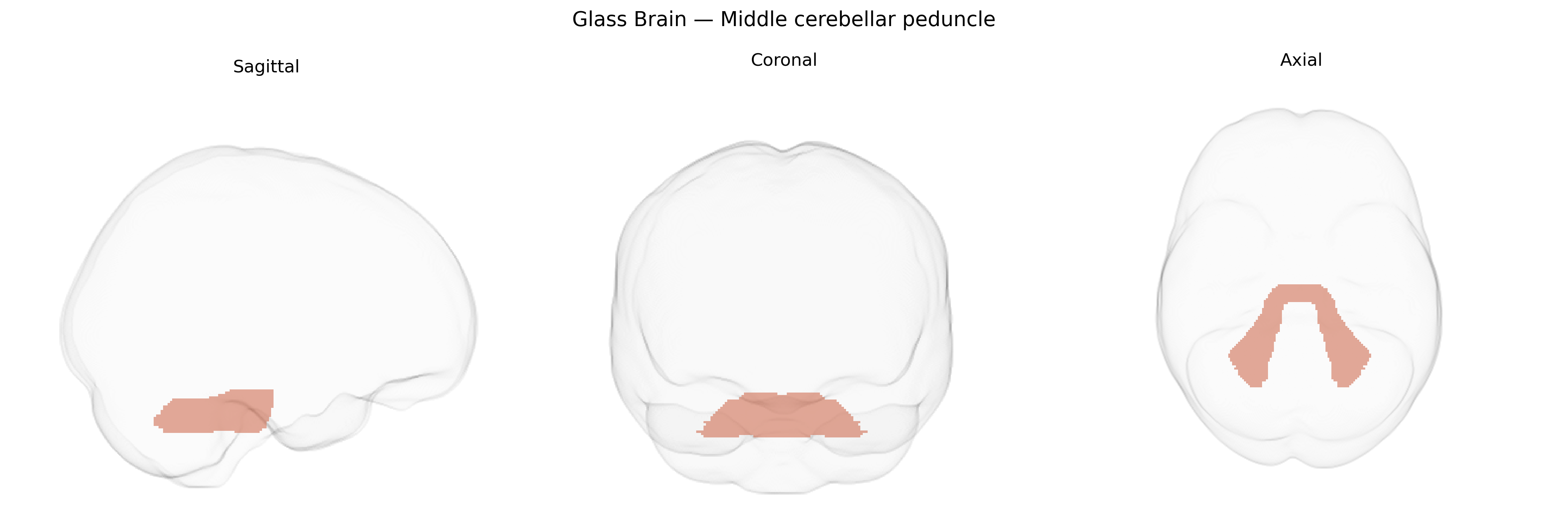
The bilateral Middle cerebellar peduncle, as defined in the JHU ICBM labels 1 mm atlas, comprises the large paired fiber bundles connecting the basilar pons with the cerebellar hemispheres. It consists predominantly of pontocerebellar fibers originating from pontine nuclei that receive input from widespread cerebral cortical regions, thereby serving as the principal conduit for corticopontocerebellar traffic involved in motor coordination, timing, and sensorimotor integration. Structurally, the middle cerebellar peduncle forms the largest of the cerebellar peduncles, occupying a lateral position between the pons and cerebellum, and is highly myelinated, making it prominent on diffusion and structural MRI. Functionally, it supports feedforward motor control and error correction by relaying processed cortical signals to the cerebellar cortex, integrating them with proprioceptive and vestibular information. Lesions in this region are associated with ataxia, dysmetria, gait disturbance, and may be involved in demyelinating or degenerative processes affecting cerebellar connectivity. Middle cerebellar peduncle
The bilateral middle cerebellar peduncle (MCP), as defined in the JHU ICBM 1 mm atlas, has been implicated in genetic studies primarily through imaging genomics and diffusion tensor imaging (DTI) GWAS that examine white matter microstructure. Large-scale GWAS of DTI measures (e.g., fractional anisotropy and mean diffusivity) have identified associations between MCP integrity and variants in genes involved in axon guidance, myelination, and neurodevelopmental processes, such as those related to oligodendrocyte function and cytoskeletal regulation, though specific loci are often shared across multiple tracts and not exclusive to the MCP. MCP abnormalities, often reflected in reduced fractional anisotropy or altered connectivity, have been linked in imaging-genetic studies to polygenic risk scores for schizophrenia, bipolar disorder, major depressive disorder, and autism spectrum disorder, supporting a role for genetic liability to psychiatric illness in shaping cerebellar–cortical connectivity. Additionally, MCP structural or diffusion alterations have been observed in carriers of pathogenic variants causing spinocerebellar ataxias and other hereditary cerebellar degeneration syndromes, indicating that genes involved in cerebellar development and synaptic signaling can impact this tract. However, there are relatively few tract-specific GWAS focused solely on the MCP, and most genetic findings derive from broader analyses of cerebellar white matter or multi-tract brain-wide association studies, where MCP emerges as one of several regions showing genetically influenced variation in microstructural properties and disease-related changes.
Overview generated by GPT-4o (2026).
Region ID: 1
Hemisphere: bilateral
Atlas: JHU ICBM labels 1mm
Full Quality Version: Download MP4
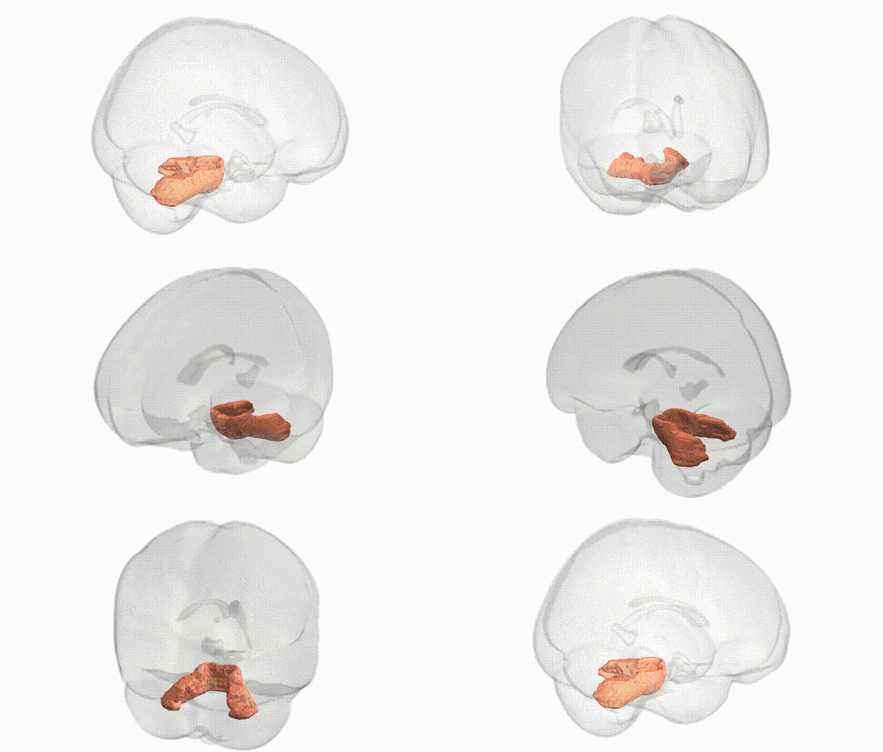
Full Quality Version: Download MP4
Wali Sidiqyar*, Gaurav Rudravaram*, Elyssa M. McMaster, Trent M. Schwartz, Adam M. Saunders, Kurt G. Schilling, Bennett A. Landman "Introducing SPINS: A Shared Public Visualization Library of Neuroanatomical Structures." Medical Imaging with Deep Learning- short paper
This resource is licensed under CC0 1.0 Universal (Public Domain).