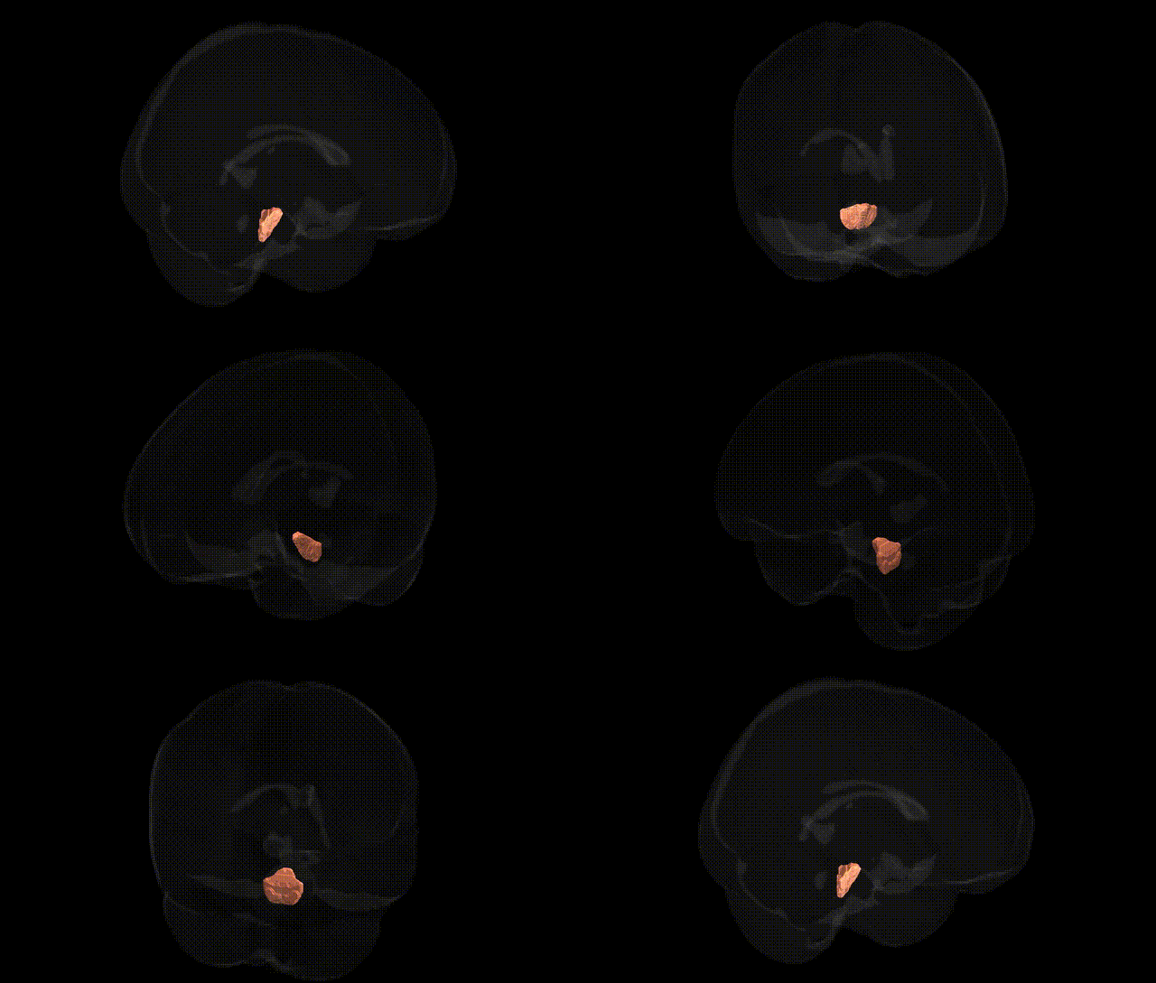
The bilateral pontine crossing tract, included within the middle cerebellar peduncle (MCP) in the JHU ICBM 1 mm atlas, comprises transverse pontine fibers that originate from pontine nuclei receiving corticopontine input and decussate to enter the contralateral cerebellar hemisphere via the MCP. These heavily myelinated fibers form a major conduit of cerebrocerebellar communication, relaying information from widespread cortical areas to the cerebellar cortex, particularly involved in coordination of voluntary movement, motor learning, and timing. Lesions affecting this tract can disrupt cortico-cerebellar loops, resulting in ataxia, dysmetria, and other cerebellar motor signs. There is no direct link for this exact tract; a related structure is the Middle cerebellar peduncle.
The bilateral pontine crossing tract, a major component of the middle cerebellar peduncle (MCP) in the JHU ICBM 1 mm atlas, has emerged in imaging–genetics and GWAS work mainly as a white-matter microstructure phenotype indexed by diffusion MRI metrics such as fractional anisotropy (FA) and mean diffusivity (MD). Large-scale studies (e.g., UK Biobank–based GWAS of diffusion measures) have identified associations between MCP/pontine crossing tract integrity and variants in genes involved in axon guidance, myelination, and neurodevelopment (including loci near or within genes such as MAG, NRXN1, CNTN4, and other cell-adhesion/axon pathfinding genes), although effects are typically polygenic and regionally pleiotropic rather than tract-specific. MCP and pontine crossing tract metrics are also repeatedly implicated in genetic studies of neurodevelopmental and psychiatric disorders—particularly schizophrenia, autism spectrum disorder, and ADHD—where risk polygenic scores correlate with altered FA/MD in this tract, suggesting shared genetic architectures influencing cortico-cerebellar connectivity. In neurodegenerative disease genetics, polygenic risk for Alzheimer’s and Parkinson’s disease has been linked to microstructural changes in cerebellar–brainstem pathways including the MCP, while rare monogenic ataxias (e.g., spinocerebellar ataxias and some POLG- or FXN-related conditions) often feature MCP/pontine tract degeneration, though typically characterized clinically or radiologically rather than via GWAS. Overall, genetic findings support a role for variants affecting oligodendrocyte function, synaptic organization, and axonal development in shaping inter-individual differences in pontine crossing tract structure, with downstream relevance for cognitive function, motor coordination, and susceptibility to neuropsychiatric and cerebellar disorders.
Overview generated by GPT-4o (2026).
Region ID: 2
Hemisphere: bilateral
Atlas: JHU ICBM labels 1mm
Full Quality Version: Download MP4
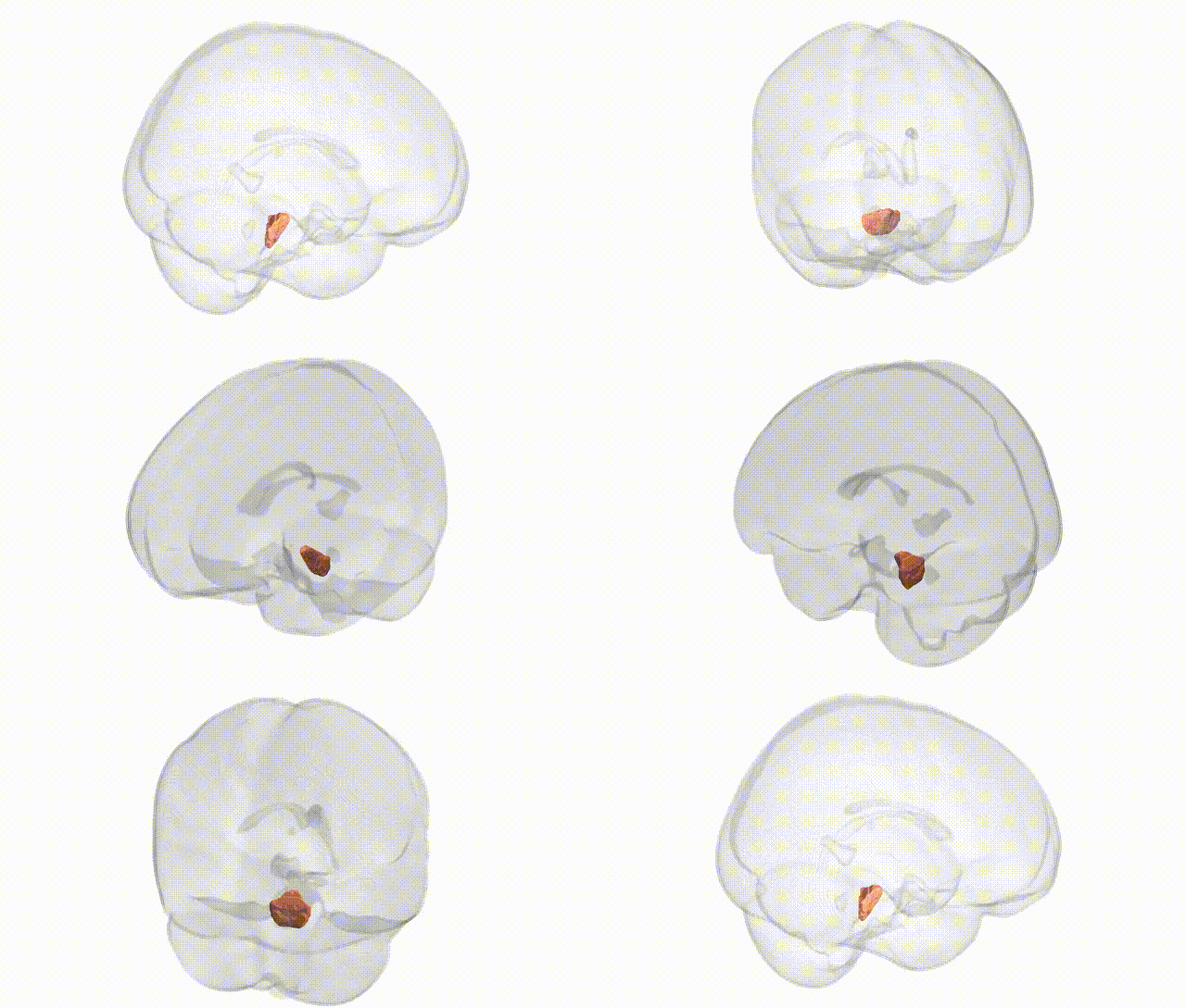
Full Quality Version: Download MP4
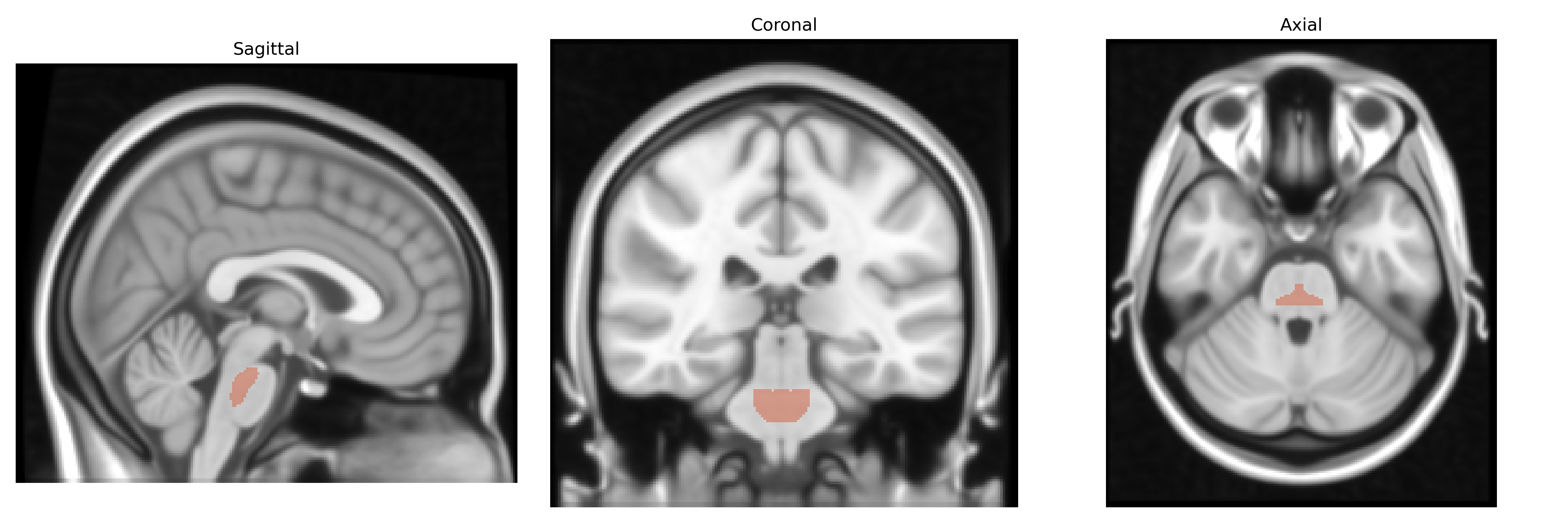
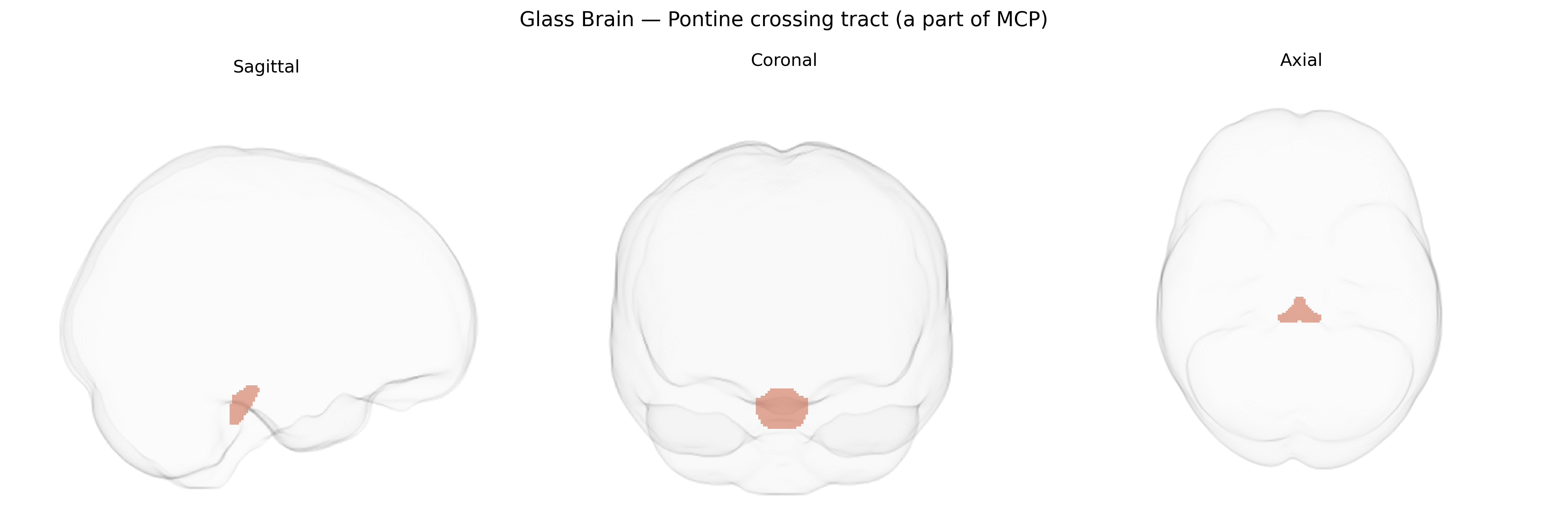
Wali Sidiqyar*, Gaurav Rudravaram*, Elyssa M. McMaster, Trent M. Schwartz, Adam M. Saunders, Kurt G. Schilling, Bennett A. Landman "Introducing SPINS: A Shared Public Visualization Library of Neuroanatomical Structures." Medical Imaging with Deep Learning- short paper
This resource is licensed under CC0 1.0 Universal (Public Domain).